
Human vision - this is one of the most important mechanisms of perception of the surrounding world. The eyes are the most mobile organs. Visually, a person receives 90% of the information. The viewing angle helps a person to navigate in the surrounding space.
Record content:
- 1 General information about the structure of the organ of vision
- 2 General operating principle
-
3 Eye structure
- 3.1 External structure of the eye
- 3.2 Internal structure of the eye
-
4 The human eye as an optical system
- 4.1 Cornea
- 4.2 Lens
- 4.3 Iris
- 4.4 Watery liquid
- 4.5 Pupil
- 4.6 Retina
- 4.7 Optic nerve
-
5 Vision angle
- 5.1 Distinctness and detail
- 6 Near and far vision
- 7 How many shades are there?
- 8 Eyes video
General information about the structure of the organ of vision
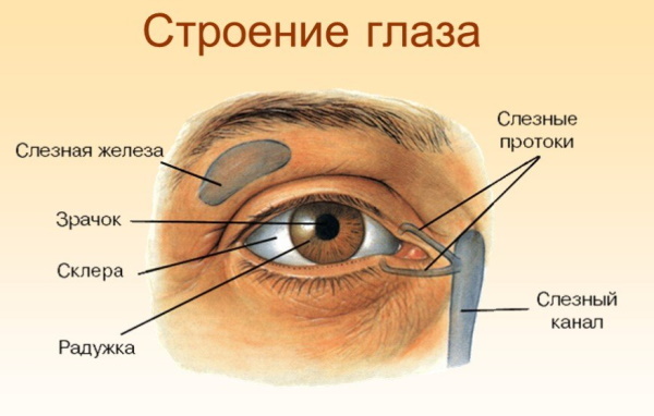
The paired visual organ absorbs visible radiation and provides visual function. The main organs of the eye are the eyeball and the optic nerve, and the auxiliary organs are the eyelids, the muscles of the eyeball and the lacrimal apparatus.
The eyes are covered with several membranes:
- protein (external);
- vascular (middle);
- mesh (internal).
The organ of vision is located in the eye sockets, protecting it from external damage. Outside, you can see no more than 1/5 of the eyeball. Most of it is in the eye socket. The eyes are spherical. The weight of each eyeball is 7 g.
General operating principle
When looking at an object, a person receives visual information. These are light rays reflected from this object, which passed through the complex optical structure of the eye and created a reduced inverted image of this object on the retina.
The information received from the retina through the optic nerve enters the brain, which allows a person to see the object as it is. The main function of the organ of vision is to transmit visual information to the brain.
Eye structure
Human eyes have an external and internal structure.
External structure of the eye
The outer parts of the eye include:
- pupil;
- iris;
- cornea;
- connective membrane (conjunctiva);
- sclera.
Internal structure of the eye
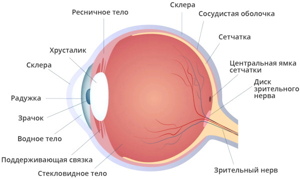
The inner parts of the eye include:
- vitreous body;
- watery liquid;
- optic nerve;
- retina;
- lens.
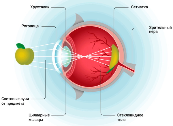
The human eye as an optical system
The human eye, which has a field of view equal to that of an F50 camera lens, is a complex optical system. consisting of cornea, iris, aqueous humor, pupil, lens, retina and visual nerve.
Cornea
The cornea is a globular and transparent part of the outer shell of the eye. With the help of thin fibrous fibers, it attaches to the ocular sclera. Due to the cornea and its structure, light waves easily penetrate into the deep layers of the human eye and end up on the retina.
The cornea has 5 functions:
- support;
- refractive;
- conductive light;
- protective.
The protective function prevents the ingress of dust and other foreign bodies. Also, the cornea immediately responds to temperature changes. With minor damage, a tear fluid is produced in the eye, which helps to get rid of the foreign body.
The cornea in a normal state has unchanged parameters. It is solid, complete, spherical, mirror-like and transparent. Its radius of curvature is 7.7-9.6 mm. The horizontal corneal diameter is 11 mm, and the refractive power is 41 diopters.
The cornea is highly sensitive, regenerative and has no capillaries.
It looks like a convex lens on the outside and concave on the inside. On the surface of the outer shell of the eye, the cornea occupies 1/5 of the part. Its thickness becomes larger along the periphery, and less in the center.

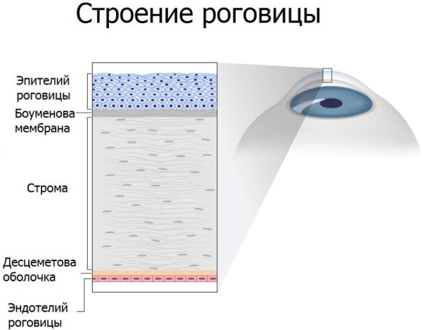
The cornea has several layers:
- integumentary or anterior, consisting of epithelial cells and is responsible for protection, gas exchange and moisture exchange;
- Bowman's or anterior borderline membraneresponsible for maintaining a spherical shape;
- stroma, formed by collagen fibers and provides resistance to damage;
- posterior boundary membrane or Descemet's sheath, contributing to the tolerance of external and internal influences on the outer layer of the eye;
- internal or endothelialcomposed of hexagonal cells and supplying all corneal membranes with nutrients from aqueous humor.
Lens
The human eye, whose viewing angle can be measured by an ophthalmologist, has a lens. It is a colorless body that is located inside the eyeball. Behind it is bordered by the vitreous, and in front - by the iris. The lens is considered a biconvex biological lens.
Its thickness varies between 3.6-5 mm. The lens diameter is 9-10 mm.
It is assembled from elongated epithelial cells that look like hexagonal prisms. Rays of light pass through the lens. In the center, this biological lens is dense. Closer to the peripheral section, it becomes thinner.
The lens consists of:
- capsules;
- capsular epithelium;
- the main substance.
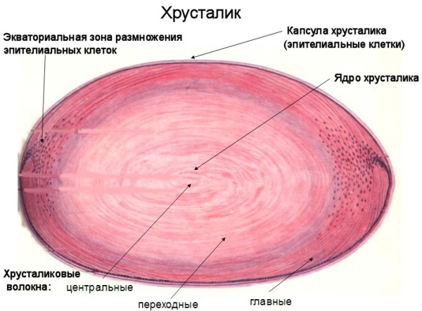
The capsule is a colorless lens shell. It is responsible for refracting light and protecting the biological lens from external negative influences. Due to the ciliary girdle, the capsule is attached to the ciliary body. On the front side, it is thicker than on the back because of the layer of epithelial cells.
The epithelium is a layer of non-keratinized cells that adhere tightly to each other and do not divide in the central zone. To the periphery, they begin to actively divide and form a zone of growth of the lens, where new fibers are formed. When new fibers appear, the old ones move to the center of the lens, forming a nucleus.
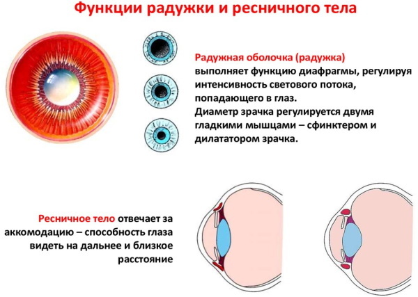
Iris
The iris is a colored disc that occupies a substantial surface of the eyeball. It is located behind the cornea in front of the lens between the anterior and posterior chambers of the eye. The required amount of light rays pass through the iris for normal vision. The thickness of the iris is 0.2 mm.
It consists of:
- anterior boundary layerformed from connective tissue cells and containing pigment cells (melanocytes);
- intermediate stromal layerconsisting of collagen fibers and permeated with a network of capillaries that carry out blood circulation;
- posterior muscle-pigment layer, which includes smooth muscles that constrict the pupil.
The first layer of the iris is subdivided into the pupillary and ciliary girdles. A circular roller is located between them. In the pupillary girdle there is a muscle that reduces the size of the pupil, and in the ciliary girdle there is a dilator - the internal eye muscle that dilates the pupil.
The supply of blood to the iris is due to several posterior and anterior ciliary arteries that form the large arterial circle. Several vessels branch off from this circle, from which a small arterial circle is obtained.
The iris is responsible for fixing the vitreous humor, evenly distributes the liquid inside the human eye, helps to focus the image on the retina and stabilizes the temperature index of the fluid in the anterior camera.
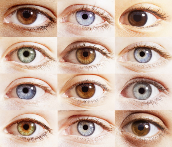
The shade of the iris depends on how many pigment cells there are. Their number is determined genetically. The iris becomes brown for the dominant trait, and blue for the recessive trait.
The color of the iris, like the pattern, changes throughout life.
At 10-12 years old, the color of the iris becomes more or less constant. In old age, its color lightens. The iris can have a specific color for one reason or another.
| Colour | Cause |
|
The content of a small amount of melanin in the iris. |
|
The content of a large amount of melanin in the iris. |
Watery liquid
The human eye, whose viewing angle depends on the size and location of the object, contains a watery liquid. It is a transparent liquid substance that fills the anterior and posterior chambers of the optic organ.
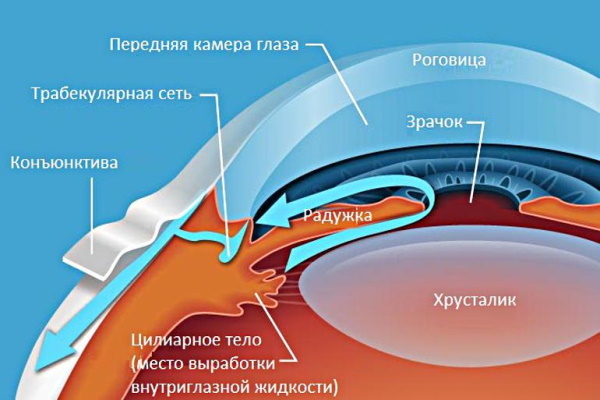
It contains nutrients such as amino acids and glucose. The watery liquid is formed by the processes of the ciliary body. The human eye produces 3-9 ml of this fluid per day.
The composition of intraocular moisture is practically the same as that of blood plasma. An exception is the amount of protein content. It is less in aqueous humor. Due to the aqueous liquid, the corneal endothelium, the lens and the anterior part of the vitreous are nourished.
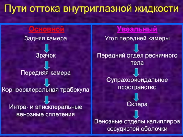
At the beginning, aqueous humor penetrates into the posterior chamber of the eye, then flows into the anterior chamber through the pupil. On the iris, this fluid rises up and then down to the cornea, flowing through the back chamber of the eye. In the corner of the anterior chamber of the human eye, an aqueous liquid is absorbed, then it passes into the Schlemm canal, and from it enters the general bloodstream.
The intraocular fluid also contains immunoglobulins. Due to them and constant circulation, there is a neutralization and removal of harmful particles from the insides of the eye. The ratio of formation and excretion of aqueous humor determines intraocular pressure.
Pupil
The pupil is a round hole in the center of the iris. Due to the ability to change the diameter, it regulates the flow of light rays that enter the eye. The diameter of the human eye can vary between 1.1-8 mm.
The degree of illumination of the retina depends on the constricting and expanding muscles of the pupil. Thanks to the autonomic nervous system, pupil size is regulated.

The dilator, which is controlled by sympathetic fibers, is responsible for the expansion of the pupil, and the sphincter, which is controlled by the parasympathetic fibers, is responsible for the reduction. Changes in the size of the human pupil occur reflexively. They depend on several factors.
Pupil dilation is carried out:
- In the dark;
- with emotional excitability;
- while feeling pain;
- when hallucinogenic and anticholinergic substances enter the body.
The constriction of the pupil begins at the moment of irritation of the trigeminal nerve and low emotional excitability.
When dim lighting changes to bright, pupil constriction occurs in 5 seconds. If bright lighting changes to dim, the pupil dilates after 5 minutes. The reaction of the pupils to light is direct or friendly.
Retina
The human eye, the viewing angle of which makes it possible to see the object in detail, is covered with the retina. It is the peripheral part of the visual organ. It is soft and inelastic.
The retina is transparent because its tissue does not contain myelin. From the outside, the retina is surrounded by the choroid, and from the inside it comes into contact with the vitreous humor.
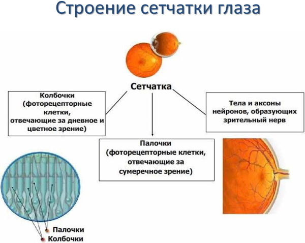
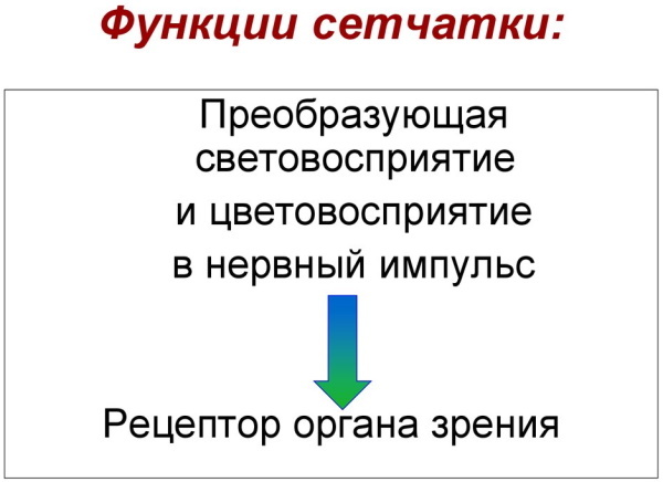
The diameter of the human retina is 22 mm. It covers approximately 72% of the inner surface of the eye. The retina is needed to convert light signals into nerve impulses. In a similar way, the primary processing of electromagnetic waves occurs. The retina has two functions: it perceives light and distinguishes colors.
The perception of light by the retina is carried out at the expense of 2 receptors - rods and cones. The receptors were so named because of their shape. Cones are divided into 3 types by color. They are red, green and blue-blue.
With their help, a person is able to recognize colors. The rods contain a pigment called rhodopsin, which absorbs the red rays of the spectrum. The rods are active at night, and the cones during the day.
In different areas of the retina, photoreceptors are distributed in an unequal number. The central region of the retina contains the most cones. The retina has 2 types of nerve cells: horizontal and amacrine. The first type of nerve cells is located in the outer plexiform layer, and the second type is located in the inner one.
The retina consists of layers:
- pigmentary, adjacent to the inner choroid and being the outer layer;
- photoreceptor, consisting of cones, rods and is responsible for color perception, as well as light perception;
- outer boundary membranethat supports the structure of the retina;
- external nuclear or granularconsisting of photoreceptor nuclei;
- external reticularpresented in the form of bipolar nerve cells, photoreceptor processes and horizontal cells;
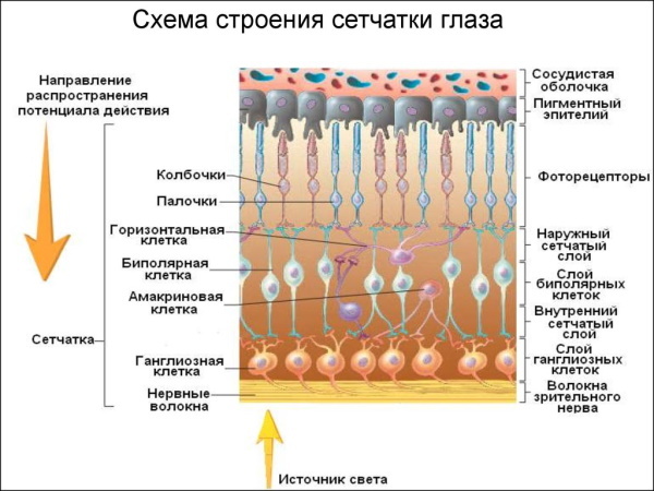
- internal nuclearcontaining the body of bipolar cells;
- inner meshconsisting of ganglion and bipolar cells;
- ganglionic, containing ganglionic multipolar cells;
- visual fiberconsisting of ganglion cell axons adjacent to the vitreous body;
- inner boundary membraneadjacent to the vitreous substance.
Optic nerve
It is a collection of fine nerve fibers. They are responsible for the transmission of primary visual impulses from retinal cells to the brain and a quick response to external stimuli.
The average length of the optic nerve is 40-55 mm. It starts from the disc - the place of accumulation of nerve cells, and ends in the chiasm - the zone in which the optic fibers are connected. Most of the optic nerve is located inside the orbit.
The optic nerve includes 4 sections:
- intratubular, representing the canal of the optic nerve;
- intraocular, being a disc with a diameter of 1.5 mm;
- intraorbital, presented in the form of an orbital part with a diameter of 3 mm;
- intracranial, which is a part of the optic nerve with a length of 1.7 cm.
The optic nerve has multiple functions
- Visual acuity. With the help of the optic nerve, human eyes are able to clearly see and examine small objects.
- Line of sight. Due to the optic nerve, the eyes see a part of the surrounding area in a motionless state.
- Color perception. The nerve helps the eyes to distinguish between the main colors and their shades.
Vision angle
The angle of view of the human eye is the total number of projections of spatial points that fall into the field of vision of a person while fixing the eye on one of several points. It allows you to quickly navigate in space. The angle of view is measured in degrees.
Its normal indicators are:
- 90 ° C - at the outer border;
- 50-55 ° C - at the upper border;
- 65 ° C - at the lower border;
- 55-60 ° C - along the inner border.
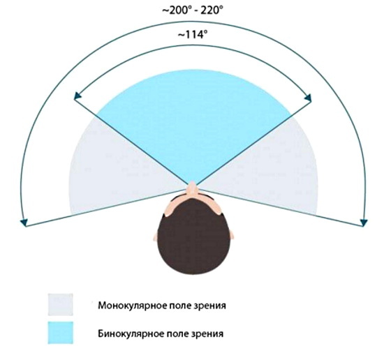
Indicators depend on:
- the structure and size of the eyeball;
- eyelid shapes;
- bone structure of the skull.
The large viewing angle makes it easier to navigate the world around. The two-eye coverage is 180 ° C horizontally and 150 ° C vertically.
Distinctness and detail
The distinguishability and detail of objects decreases depending on the distance from the central object on which the vision is focused. The farther the subject is from the focus area, the less detail and clarity it becomes.
Near and far vision
Human eyes can see objects both near and far. Accommodation is the ability to see objects clearly at different distances. The brighter the object, the better it can be seen at long distances. The main thing in this ability is the elasticity of the lens.
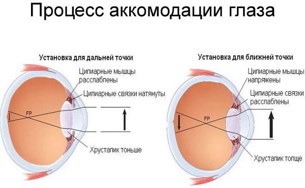
The lens in its normal state can change its shape and adapt to near or far vision, depending on the selected object. The flat and elongated lens allows you to distinguish between objects in the distance. The lens takes on a hemispherical shape when a person looks at nearby objects.
The area of accommodation is the distance between the nearest and farthest points of clear vision. When considering distant objects in people with farsightedness, the ciliary muscles are strained - the internal paired muscle of the eye, which provides accommodation.
People suffering from myopia have insufficiently developed accommodation. They can only see well at short distances.
How many shades are there?
Human eyes normally distinguish 4 primary colors. They are red, green, yellow and blue. By merging the primary colors, different shades are obtained. The human eye is capable of recognizing 100 shades.
The human eye is complex enough. Its constituents perform various functions that are necessary for vision. The viewing angle depends on many factors and can change with diseases of the optic organ.
Eyes video
Vision documentary:


