
The back wall of the eye differs in a complex structure, but histology reveals even minor deviations. The individual parts of the organ are closely interconnected, often consist of several membranes, and have additional structures. If even one of them malfunctions, vision deteriorates. If treatment is not started on time, a person may go blind.
Record content:
-
1 Basic functions of the eye
- 1.1 Structure
- 1.2 Cornea
- 1.3 Anterior chamber of the eye
- 1.4 Iris
- 1.5 Pupil
- 1.6 Lens
- 1.7 Vitreous
- 1.8 Retina
- 1.9 Sclera
- 1.10 Choroid
- 1.11 Optic nerve
- 2 Violations
- 3 Video about the structure of the eye
Basic functions of the eye
The main function of the eyes is visual perception of the environment. The organ catches the reflection of light rays from objects. It penetrates the pupil (the lumen in the iris), the cornea, refracts and concentrates in the lens (this is a capsule with liquid). The radius of curvature is changed with the help of muscles. Distant objects appear flatter, and nearby objects appear convex.
This process is called accommodation, and the streams of light are focused on the retina. In its receptors, light energy is transformed into electrical energy and transmitted to visual neurons. Further, the information moves along the nerves to the brain, where decoding, analysis begins, and the person receives a visual picture.
Structure
Normally, an organ in an adult is a slightly flattened sphere with a diameter of 25 mm. In newborns, the weight of the eyeball is up to 3 g, in adults - up to 8 g. Inside - structures, tissues, with the help of which all images of the environment are transmitted to the brain.
The eye consists of three key systems:
- the central one is located in the cerebral cortex, where information is evaluated and visual images are formed;
- conductive, which consists of nerve fibers;
- peripheral - the eyeball and nearby tissues.
With the help of a conductive system, the perception of external images is transmitted to the brain, where information is processed.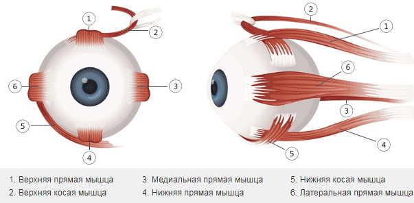
| Parts of the eye | |
| External | Internal |
|
|
The outer parts protect the eye from damage, its surface must always remain moist. The internal structures of the organ are needed to refract light rays. Without this, the retina will be damaged, causing complete blindness.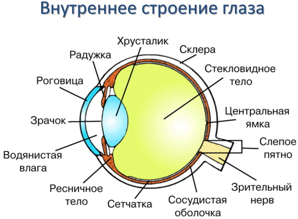
The structure of the eye includes auxiliary components:
- eye sockets;
- lacrimal fluid production apparatus;
- conjunctiva;
- eyelids;
- oculomotor muscles.
The back wall of the eye also contains structures that provide life support - blood supply, regulating the production of tears, hemo- and hydrodynamics. When checking for histology, 2 integumentary membranes are assessed (conjunctiva, bottom of the apple) and 3 main ones - vascular, reticular, fibrous.
The largest parts of the organ are the vitreous body, the lens. The eyelids are folds of skin that protect the eye from external irritants. Also, a secret comes out through them, which reduces the friction of the shells, preventing their damage.
Around the apple are muscles that are associated with the eyelids, due to this, blinking is carried out. The internal structures also have muscles that are needed to change the size of the pupil, the shape of the lens.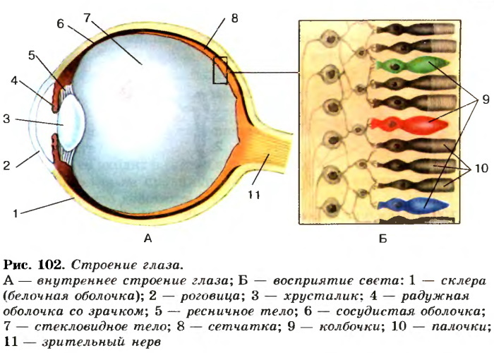
An important separate structure is the lacrimal canal. It consists of glands with fluid secretion, placed in a sac. Excess fluid is removed through a special channel. It not only moisturizes the eyes, preventing them from drying out, but also has an antibacterial effect, preventing the spread of dirt and pathogenic bacteria into the internal structures.
Cornea
The posterior wall of the eye (histology describes the anatomy in detail) is closed from direct mechanical action. There are many vulnerable structures there. For example, the cornea is a transparent, homogeneous part of the fibrous layer. It is thinner in the middle than at the edges. Spherical, with a mirror surface, without blood vessels. The cornea has high pain and tactile sensitivity. It protects the eyes from the outside.
The place where the cornea smoothly passes into the sclera is called the limbus (it is an almost transparent ring up to 1 mm wide). Light rays penetrate through the cornea, and a person perceives a three-dimensional image. There are no blood vessels in it.
It consists of layers:
- Epithelial. It is located outside the cornea. The epithelial layer regulates the level of moisture from the lacrimal glands. Oxygenation of cells occurs through the tear film. The integrity of the cornea is disturbed by the ingress of foreign bodies into the eye - debris, dust, solid particles. But if they did not penetrate deeply, then the damaged epithelial cells quickly regenerate without pain.
- Bowman's membrane. This is also the superficial layer under the epithelial layer. But unlike him, it is not restored, and any damage to it impairs vision. The function of the membrane is to nourish the cornea, to participate in cellular metabolism.
- Endothelial. It is responsible for removing excess fluid. This layer is poorly regenerated. Normally, its density is 1500-3500 cells / 1 mm², and with age it decreases and gradually loses its functionality. After injury or severe inflammation, the number of cells decreases to 800 or less. The cornea swells, and the clarity of vision is reduced.
- Stroma. It is a layer of collagen fibers that fill the empty space.
- Descement membrane. It is a thin membrane at the border of the stroma that separates it from the endothelial mass.
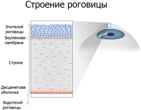 The last layer of the cornea is the tear film. It softens fabrics, moisturizes and cleanses. The lacrimal fluid washes away dirt and dust, ensures normal oxygen permeability.
Anterior chamber of the eye
The posterior wall of the eye (histology allows you to evaluate not only parts of the organ externally, but also to check the cavities) has two chambers with fluid. The front is the space behind the back of the cornea.
It is located directly in front of the iris, near the middle of the lens. The rear camera is behind the iris. Limited on both sides by the vitreous, ciliary body. In the outer part of the anterior chamber, there is a drainage system made of a mesh, collector tubules, and scleral sinus.
The cameras communicate with each other through the pupil. In both, fluid is continuously circulating, saturating the eye with vitamins, minerals and nutrients. The result is increased metabolism and rapid tissue regeneration. Also, the liquid in the chambers is needed to refract the rays of light. When the balance between the chambers is disturbed, intraocular pressure increases.
Iris
The iris is located behind the anterior chamber. Their color is largely dependent on pigmentation. In newborns, its synthesis has not yet been regulated, so more often children are born with light blue eyes.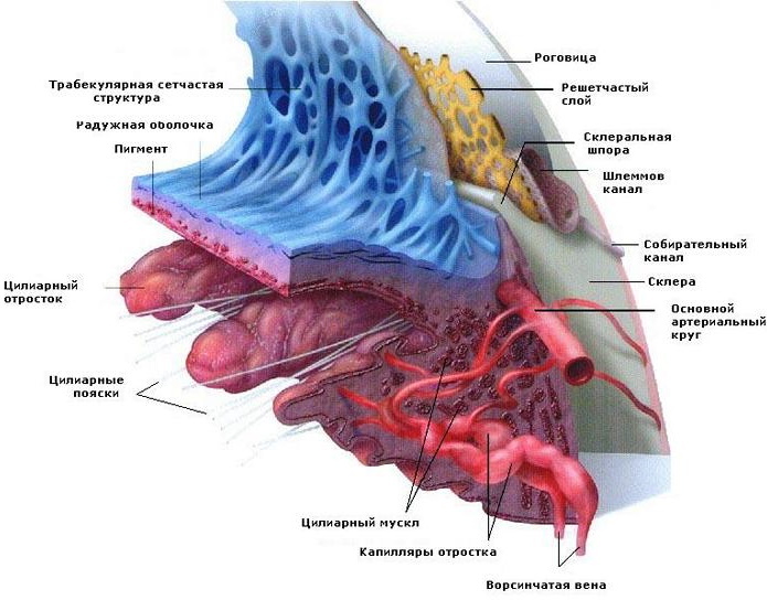
But as they grow older, they can change color. The iris consists of muscle fibers that are able to instantly relax and contract. Thus, the luminous flux is regulated and the size of the passage channel changes.
Pupil
In the middle of the iris is the pupil. Under muscular influence, its diameter changes - depending on the lighting. The more it is, the smaller the pupil clearance. When a person is in the dark, the muscles tense. The pupil expands, allowing you to see the surrounding objects.
Its normal work is disrupted by diseases or under the influence of drugs. The pupil's response to light is important in the diagnosis of the eyeball. But prolonged exposure to light flux can impair visual perception.
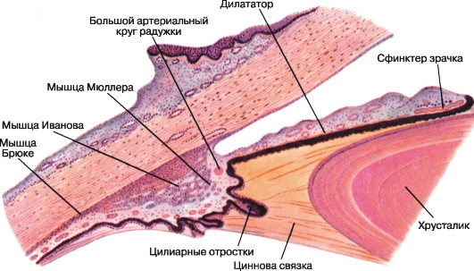
Lens
The lens is responsible for the clarity of vision, focusing. It is a transparent semi-solid lens, convex on both sides, which is held in place by the ciliary band. The lens diameter is 9-10 mm, the thickness is 3.5-5 mm. In infants, it is spherical, softer in structure.
The lens is located in the vitreous fossa, behind the iris, and is held in place by fibers. The back of the organ is washed with moisture through a small gap, since there is a small free space. On the outer edge of the fossa is the annular Viger ligament.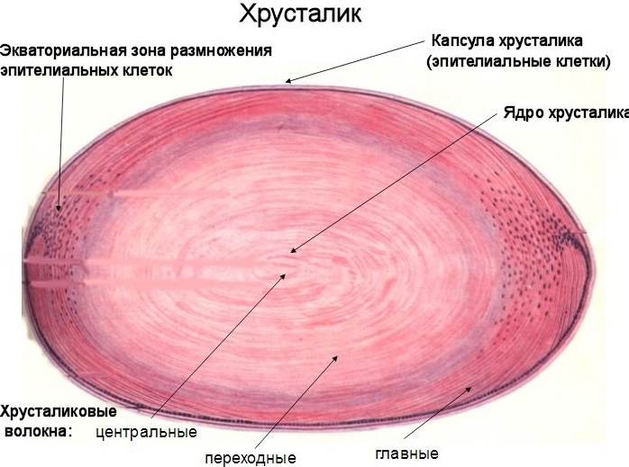
The lens is highly elastic and instantly takes the desired shape to adjust the clarity of the outlines of objects at different distances. With age or due to illness, it becomes cloudy, cataracts develop, and the sharpness of vision is impaired. It can be restored by replacing the lens with an implant.
Vitreous
The vitreous humor is a dense, gelatinous gel that maintains the shape of an apple. Fills the empty space inside it. The volume of the vitreous body is 3.5-4 ml, and the mass is 4 g. it is surrounded by a protective membrane.
On the one hand, it joins the lens, on the other, to the retina. The body has two main components. The vitreous stroma is made of collagen fibrils and loose matter with small areas of void that are filled with fluid. The second component is the hyaloid membrane.
The vitreous body evens out intraocular pressure after surges, eliminating the negative consequences after sudden changes. Transparent walls redirect light rays to the retina, and a complete picture of what you see is created.
Retina
The back wall of the eye (histology allows you to pay attention to especially vulnerable structures) is covered with the retina. It lines the vascular layer. It is well fixed near the optic nerve, along the border of the macula, at the dentate line, but in other areas it quickly exfoliates.
The thickness of the retina varies depending on the location, has 2 parts - cilio-iris, optical. The second is a highly differentiated tissue with photocells - cones, rods.
They help to recognize the color palette:
- Cones help you see details. They need good lighting to perceive light waves. Then the eye can distinguish any shades, consider even very small elements.
- The sticks are highly sensitive. Thanks to them, a person sees even in low light, when the subject is out of focus. Sticks are needed for lateral vision, panoramic vision.
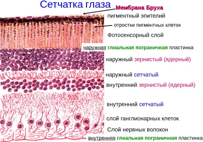
The structure of the retina histology
Photocells perceive rays of light 380-770 nm long. The optical part is located between the nerve disc and the ciliary body, consists of ten layers. The closer to the macula, the more the retinal anatomy changes - nerve fibers and some layers disappear.
The nerve disc is located in the nasal part of the retina. There are no photoreceptors, there is a blind area. The main blood vessels of the retina are located in the thickness of the nerve fibers, forming a capillary network (it is absent in the macular region).
Sclera
The sclera is an opaque dorsal membrane (0.3-1 mm) in the apple, which faces the orbit. In the area of the optic nerve, it forms a lattice. The thinnest areas of the sclera are very vulnerable to injury and high intraocular pressure. It connects to the cornea and conjunctiva of the apple near the limbus. It is surrounded by a dense nerve plexus.
On the anterior edge of the sclera, on the inner side, there is a circular groove - up to 0.75 mm wide. Its rear edge is curved like a spur. The ciliary body is attached to it. On one side, the groove is bordered by the cornea, the venous canal is located at the bottom, and the rest is in the form of a mesh.
There are few vessels and nerve endings in the sclera. Six motor muscles are attached to it, which regulate the position of the apple. The structure of the sclera is much denser than the cornea, as it is responsible for supporting the shape of the apple and its movements. The sclera prevents the penetration of light inside, protecting the eyes from the inside. There are nerve endings, vessels that feed the eye.
Choroid
The surface of the sclera is covered with a choroid (choroid), which provides blood flow. There are no nerve endings that could notify painful sensations of disturbances in the work of this area. The choroid does not interact with the ciliary arteries in front. It is partially associated with the retina, therefore, in diseases, pathological changes affect it as well.
The ciliary body, which occupies the middle 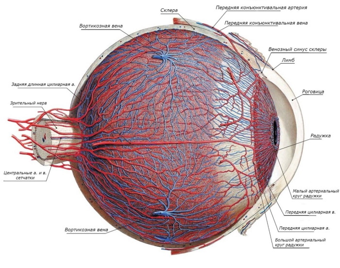 area of the choroid. It borders on the sclera with a 6-7 mm girdle. Inside, the ciliary body is in contact with the lens. The vasculature is formed by small arteries passing near the sclera and peels off with ease.
Optic nerve
The optic nerve consists of a disc, nerve fibers, chiasm (this is the place where nerve trunks intersect) and a section where it passes into the brain. Nerve fibers, 5-6 cm long, begin at the retina. Then they connect in the brain to form chiasm. Then the fibers are pulled to the main center, where the signals are decoded.
The optic nerve is covered by three sheaths, different in structure - soft, cobweb-like and hard. The thickness of the organ without them is 3-3.5 mm, if with them, then 4-4.5 mm. The nerve grows together with the periosteum. Its intracranial part and chiasm are covered only by a soft membrane. All nerve fibers are collected in 3 bundles, but the organ itself is divided into two parts - left and right, there are no sensory endings.
Violations
The posterior wall of the eye (histology helps to select the desired site for examination) is the most vulnerable part of the organ. Visual impairment can be congenital or acquired.
In the second case, this often happens at school, when the load on the eyes increases greatly, and children do not keep distance when reading, do not pay attention to lighting when doing on their own lessons. But more often visual impairment occurs in 50 percent of adults.
The most common problems are myopia, hyperopia, or a combination of both. The main reason is the abnormal structure of the eyes or ophthalmic diseases. With farsightedness, a person does not distinguish between objects and the surrounding near, but well - those that are far away.
More often, the cause is age-related changes after 45 years, which gradually intensify. Farsightedness develops due to a decrease in the eyeball, lens or its displacement, flat cornea.
With myopia, a person sees well up close, and the further the object is, the more it diffuses. More often, this is a hereditary disease or develops in schoolchildren when the load on the eyes increases or due to excessive curvature of the cornea. With myopia, the structure of the eye changes - the eyeball becomes larger, the image falls not on the retina, but in front of it.
The back wall of the eye is clearly visible using modern devices. If abnormalities are found during histology, then more often they can be eliminated with the help of glasses. In difficult cases, surgery is required (for example, lens replacement). If even extreme measures do not help, then the person becomes blind.
Video about the structure of the eye
Anatomy of the eye:


