Content
- Type F-I (active bleeding)
- I a (pulsating jet)
- First aid for gastroduodenal bleeding type F-Ia
- Medication to stop pulsating gastric bleeding
- Clipping
- Surgery
- I b (bleeding)
- First aid for gastroduodenal bleeding type F-Ib
- Electrocoagulation of the vessel
- Argon plasma coagulation
- Type F-II (signs of recent bleeding)
- II a (visible, non-bleeding vessel)
- First aid for gastroduodenal bleeding type F-IIa
- II b (fixed thrombus-clot)
- First aid for gastroduodenal bleeding type FII-b
- II c (flat black spot, black bottom of the ulcer)
- First aid for gastroduodenal bleeding type F-IIc
- Type F-III (ulcer with a clear, white bottom)
- Bleeding Videos
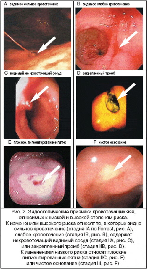
Classification of bleeding by J. A. Forestu was approved in 1974. This is a unified medical system that is used by endoscopist doctors in the process of diagnosing patients with signs of stomach ulcers, as well as damage to the mucous membrane of the duodenum. This classification method allows us to unify the general type of gastroduodenal ulcer, exclude the factor of discrepancies, and predict the potential risk of recurrent internal bleeding.
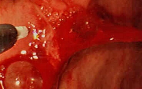
Type F-I (active bleeding)
Forest's classification of gastroduodenal bleeding includes 3 stages of ulcerative lesions of the gastrointestinal tract, accompanied by the release of blood. During the diagnosis, the doctor performing endoscopy evaluates the appearance of the ulcer, the rate of blood flow, its color shade, the rate of formation of a dense thrombus. These indicators form the basis for further prognosis of positive or negative dynamics to the patient's recovery.
I a (pulsating jet)
Active gastroduodenal bleeding of type F-Ia is characterized by a jet discharge of gushing blood. In the process of diagnosing a patient, an endoscopist doctor finds a damaged arterial vessel. The patient's stomach is rapidly filled with blood, which is distinguished by a light scarlet hue. A pulsating stream of gastroduodenal bleeding is a pathological sign, the elimination of which requires the provision of emergency medical care to the patient.
Acute internal blood loss of type F-Ia according to Forest's classifier is accompanied by the following additional symptoms:
- lowering blood pressure;
- dizziness;
- violation of coordination of movements;
- Strong headache;
- darkening in the eyes;
- noise in ears;
- acute pain in the abdominal cavity, which is localized in the upper central part of the abdomen;
- growing anemia;
- the release of fresh blood from the rectum;
- bloody vomiting.
In especially severe cases with extensive damage to the arterial vessel, pulsating gastroduodenal bleeding causes the patient to lose consciousness. There is a risk of death.
First aid for gastroduodenal bleeding type F-Ia
The Forest classification of bleeding systematizes the clinical cases of ulcerative bleeding in patients with chronic gastrointestinal diseases. In the presence of gastroduodenal blood loss of type F-Ia, the patient needs emergency medical care.
The table below lists the mandatory rules of endoscopic tactics that are followed in the diagnosis of patients with gushing gastric bleeding.
| Endoscopic Tactics Rules | Doctor's actions |
| 1. Determination of the source of jet bleeding. | At this stage, the primary task of the doctor is to detect the focus of arterial bleeding. This stage of the diagnostic process should last a minimum amount of time. Delay is fraught with the development of severe complications and a further deterioration in the general well-being of the patient. |
| 2. Diagnostic assessment of the nature of bleeding. | After finding the source of acute blood loss, the endoscopist must urgently assess the intensity of gastroduodenal bleeding. At this stage of the examination, the doctor determines whether the flow of blood into the stomach is constant, lasting, or whether the physiological mechanism of thrombus formation is running. |
| 3. Endoscopic hemostasis. | The final stage of endoscopic tactics for providing first aid is the immediate arrest of internal bleeding. The doctor independently decides which method of hemostasis to apply in a specific clinical situation. |
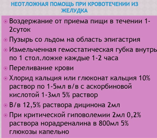 Type F-Ia endoscopic stopping of gastric bleeding involves the use of injection hemostasis, the imposition of metal staples, and a surgical operation.
Medication to stop pulsating gastric bleeding
One of the most common methods for stopping gastroduodenal bleeding of type F-Ia is the injection of a hemostatic agent. The success rate of this therapeutic tactic is 70-100%.
In the process of providing first aid to the patient, the endoscopist performs the following actions:
- Directs endoscopic equipment to the source of arterial bleeding.
- Injects the stomach tissue located in the circumference of the damaged vessel.
- Fixes the result of therapeutic manipulations.
With a positive therapeutic effect, the patient should have a gradual cessation of gastric bleeding. The constituents of drugs that are injected into the tissues near the injured vessel have vasoconstrictor properties.
In addition to this, stopping gastroduodenal bleeding is achieved by mechanical compression of the walls of the destroyed artery. Strengthening natural thrombus formation accelerates the process of complete relief of acute blood loss. Despite the availability, simplicity, and effectiveness of injectable stopping of type F-Ia gastroduodenal bleeding, the risk of ulcer reopening remains. The likelihood of recurrence is 25%.
Clipping
The therapeutic method for clipping gastric bleeding type F-Ia involves the use of more invasive therapeutic procedures. In this case, the endoscopist applies metal braces to the walls of the damaged vessel or tissue in the area of the focus of acute blood loss. Special endoscopic equipment is used in the form of a clipper inserted into the stomach cavity through the instrument tube of the endoscope.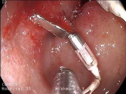
The use of this method is indicated for patients diagnosed with gushing gastric bleeding with extensive damage to the arterial vessel. Clipping is effective in cases where the injection of hemostatic agents has not brought a positive result.
Surgery
The use of the method of surgical treatment is indicated for patients with extensive gastric bleeding from an arterial vessel. For example, if, according to the results of endoscopy, it was determined that the use of injections and clipping would not be able to provide a complete cessation of acute blood loss.
Surgical elimination of gastroduodenal bleeding of type F-Ia is carried out according to the following algorithm of actions:
- The patient is urgently admitted to the hospital of the surgical department.
- After preliminary preparation, the patient is transported to the operating room.
- The anesthesiologist puts the patient into a state of medication sleep. The dosage of drugs that provide general anesthesia is selected on an individual basis.
- The surgeon makes an incision in the patient's abdomen, gaining access to the source of the bleeding.
- With the help of surgical instruments, the walls of the damaged arterial vessel are sutured, and the adjacent tissues of the stomach are sanitized.
- After stopping the bleeding, the surgeon closes the patient's abdominal cavity using suture material.
In the first 3 days after this operation, the patient is in the hospital of the surgical department in the intensive care unit. In the absence of recurrent bleeding and signs of complications, the patient receives further treatment antibacterial medicines, medicines that normalize the secretory function stomach.
I b (bleeding)
The Forest classification of bleeding is a unified system for assessing pathological conditions of the gastrointestinal tract. Gastroduodenal blood loss type F-Ib is considered a less dangerous complication of peptic ulcer disease. In the presence of this pathology in a patient undergoing endoscopic examination of the stomach, venous bleeding of low intensity is found. Leakage of blood into the stomach cavity occurs constantly or during an exacerbation of peptic ulcer disease.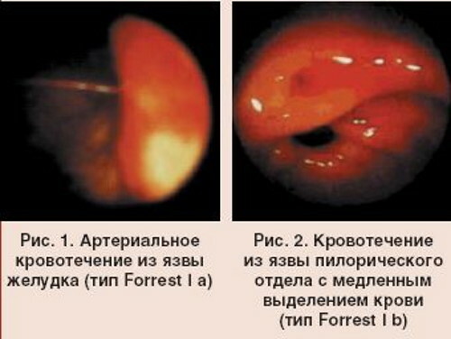
The subject is diagnosed with the following additional symptoms:
- aching or cramping pain in the upper center of the abdominal cavity;
- periodic dizziness;
- darkening of feces;
- a decrease in the level of hemoglobin in the blood;
- the development of anemia;
- pallor of the skin;
- decreased appetite;
- nausea.
According to the results of an endoscopic examination of the patient, it is found that traces of venous blood are present inside his stomach, the particles of which look like dark flakes. In patients with gastroduodenal bleeding type F-Ib, periodically vomiting occurs with the release of a homogeneous mass in the form of coffee grounds. This is a consequence of low intensity parenchymal bleeding.
First aid for gastroduodenal bleeding type F-Ib
For patients with this type of gastric bleeding, injection of hemostatic drugs, as well as the imposition of metal braces, are indicated. Depending on the volume of blood secreted, it is possible to use innovative methods of electrocoagulation and argon plasma coagulation of the damaged vessel. These methods of treating type F-Ib gastroduodenal bleeding are effective in the presence of mild bleeding.
Electrocoagulation of the vessel
Stopping gastric bleeding using electrocoagulation is carried out under sterile conditions in an endoscopic diagnostic room.
This method of therapy is carried out according to the following algorithm of actions:
- The patient lies on his side with endoscopic equipment inserted into his digestive tract.
- An electrocoagulator is directed through the instrumental canal in the area of localization of low-intensity gastric bleeding.
- By the action of an electric current, the doctor cauterizes the walls of the damaged vessels.
- Upon completion of the therapeutic manipulations, the electrocoagulation equipment is removed from the patient's digestive system through the instrument channel of the endoscope.
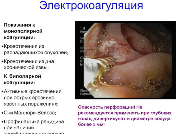
A positive therapeutic result is achieved through thermal contraction of the collagen particles that form the vascular wall. Due to this effect, a narrowing of the lumen in the damaged vessel is provided with further formation of fibrin. The risk of recurrent rebleeding is minimal. The formation of a dense thrombus guarantees gradual healing of the ulcer formation.
Argon plasma coagulation
It is believed that the method of argon plasma coagulation of gastroduodenal bleeding type F-Ib allows to achieve maximum therapeutic efficacy in the treatment of complications of gastric ulcer. The principle of this technique is to cauterize the walls of a blood vessel with a flow of argon plasma.
The advantage of this therapeutic direction over other methods of coagulation is that the doctor gets the opportunity to arrest pathological conditions of tissues to a depth of 3 mm.
The method of argon plasma coagulation is effective not only for superficial bleeding of low intensity, but also for deeper damage to the walls of the stomach. These therapeutic manipulations can be performed immediately after the endoscopic examination of the gastrointestinal tract is completed.
Standard coagulation of the vessel walls with electric current allows the doctor to operate only in the axial direction. This limits the therapeutic options for stopping superficial bleeding. Cauterization of damaged tissues with a flow of argon plasma can be carried out in the axial, radial and transverse directions.
Type F-II (signs of recent bleeding)
The Forest classification of bleeding is a scale for assessing complications that arose during the next exacerbation of peptic ulcer disease. Signs of recent gastrointestinal bleeding are fixed in a separate type F-II.
II a (visible, non-bleeding vessel)
A distinctive feature of the consequences of gastroduodenal bleeding of type F-IIa is the preservation of fresh signs of recent blood loss. In the process of endoscopic examination of the patient, it is found that a thrombosed vessel with signs of traumatic injury is present in the pathological focus. The appearance of this symptomatology is the result of sluggish venous bleeding.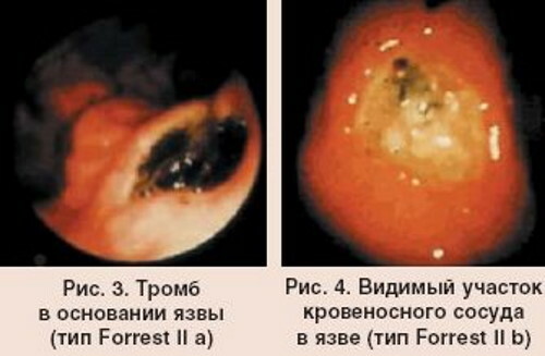
In a patient with similar symptoms, the following additional symptoms of a pathological condition of the gastrointestinal tract are present:
- nausea;
- cramping pain in the stomach;
- vomit;
- physical weakness;
- loss of appetite.
Damage to the walls of the digestive tract with the presence of a visible vessel without the release of blood is dangerous with a recurrence of gastroduodenal bleeding. The likelihood of repeated blood loss exceeds 50%.
First aid for gastroduodenal bleeding type F-IIa
Patients with a thrombosed vessel in damaged areas of the gastrointestinal tract do not need surgical treatment. Prevention of recurrence of internal bleeding is achieved by clipping destroyed tissues, squeezing the walls of the damaged vessel with injectable drugs with hemostatic properties.
II b (fixed thrombus-clot)
The Forest classification of bleeding simplifies the process of diagnosing emergency conditions in patients with complications of ulcerative lesions of the digestive tract. The presence of a fixed thrombus or a dense blood clot in the structure of damaged tissues indicates a slow restoration of the integrity of the vessel walls.
In this case, the patient continues to feel general malaise, nausea, loss of appetite, and bouts of aching pain. The appearance of a fixed thrombus-clot in the ulcer is a positive sign of early tissue healing.
First aid for gastroduodenal bleeding type FII-b
Patients with type FII-b gastroduodenal bleeding do not need emergency medical attention. In the presence of a dense fixed thrombus, the attending physician prescribes the patient further pharmacological therapy that reduces the level of acidity of gastric juice, and also protects the walls of the gastrointestinal tract from further damage.
Endoscopic observation of the rate of tissue healing is performed periodically. In this case, the use of electrical and plasma coagulation is not advisable, since the walls of the blood vessel are already closed by the formed high-density thrombus. Clipping of the ulcer will cause additional damage to the gastrointestinal tract.
II c (flat black spot, black bottom of the ulcer)
The consequence of gastroduodenal bleeding according to type F-IIc has signs of a borderline state, in which in the outbreak pathology of the digestive tract is no longer a fixed blood clot, but there are still signs of ulcerative education.
Such symptoms indicate a positive trend towards the restoration of the damaged vessel, but the risk of recurrence of internal bleeding is still at the level of 15-20%. In the process of endoscopic examination of the stomach or the walls of the duodenum of the patient, a black bottom of the ulcer with traces of hemorrhagic impregnation is found.
First aid for gastroduodenal bleeding type F-IIc
In patients with this diagnosis, there are no signs of acute or flaccid bleeding into the gastrointestinal tract. To minimize the risk of ulcer reopening, the patient is prescribed restorative pharmacological therapy.
The attending physician selects medications that reduce the acidity of gastric juice, envelop its walls from the destructive effects of digestive enzymes. Bismuth-based drugs can be used to accelerate the regeneration of damaged tissues.
Type F-III (ulcer with a clear, white bottom)
The consequence of gastroduodenal bleeding of type F-III provides for the cleansing of the bottom of the ulcer from signs of hemorrhagic impregnation. According to the results of endoscopic diagnosis of the patient, there are no signs of bleeding at all. At this stage, the risk of reopening arterial or venous blood loss is minimal. In the absence of the negative impact of pathological factors, further tissue scarring continues.
The classification of bleeding according to Dr. Forest is an international system for assessing signs of a pathological condition of the digestive tract in patients with peptic ulcer disease. This technique is used by endoscopic doctors. The advantage of the Forest classifier is the simplicity and versatility of this diagnostic method.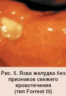
The most dangerous gastroduodenal bleeding is considered to be a gushing discharge of blood from an arterial vessel. Treatment of this pathology requires an urgent surgical operation to restore the integrity of the damaged vessel. To stop venous bleeding of low intensity, use the technique of clipping, injection compression of tissues, as well as their cauterization by electrocoagulation.
Bleeding Videos
Gastrointestinal bleeding: