Content
- Features of the structure of the muscles of the legs
- Separation of leg muscles
- Flexor and extensor muscles of the leg
- Pelvic musculature
- Front group
- Posterior-outer group
- Thigh muscles
- Front group
- Medial group
- Posterior muscle group
- Calf muscles
- Front group
- Lateral group
- Back group
- Muscles of the foot
- Rear group
- Medial group
- Lateral group
- Middle group
- Videos about leg muscles
Muscles account for about 50% of a person's body weight. Muscle legs - the most extensive, varied and numerous of such fibers. In the anatomical atlas, you can see color photos with a detailed description of the structure, biomechanical characteristics and location of the muscle groups of the lower extremities.
Features of the structure of the muscles of the legs
Elastic, dense and durable fibers take the load of the whole body, provide mobility when walking, running, jumping. The functional purpose of the muscle groups of the legs is reflected in the features of the anatomical structure of the muscles.
Each organic tissue of this type is attached to tendons, which provide anchoring to skeletal structures or small bone formations. The structural features of the muscles of the lower extremities determine increased endurance, resistance to mechanical stress.
The muscles of the legs of a person in the photo with the description are often shown in section. This allows you to study the internal structure of muscle fibers. An important feature of the anatomical structure of the muscles of the lower extremities is better development in comparison with the muscles of most other parts of the body.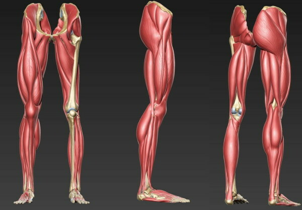
Evolutionarily, such a structure is due to the need to constantly be in an upright position. During physical activity, the muscles of the legs consume a large amount of energy resources. Well-developed musculature of the lower extremities reduces the load on the more fragile and deformable spine.
Separation of leg muscles
Such groups of organic fibers are classified according to their location, functional purpose, and biomechanical characteristics.
According to the zonal distribution of the leg muscles, they are divided into the following groups:
- Pelvic girdle. Such muscles do not belong to the anatomy of the lower extremities, but they are attached to the femoral musculature and, when moving, work in conjunction with it. The complex of the pelvic girdle is localized mainly in the back of the body.
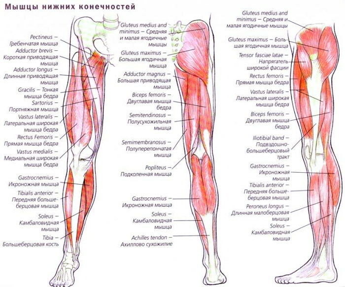
- Thigh muscles. These muscles determine the shape of the legs. Hip groups originate from the pelvic joint and extend to the knee joint.
- Shin muscles. Not the largest, but extremely hardy muscles. They extend to the area of the leg between the knee and the foot, providing anatomical interconnection of the fibers located in these zones. The lower leg muscles are divided into anterior, medial and internal groups.
- Muscles of the foot. They perform a support function, ensuring the stability of the body in an upright position.
- Small muscles of the fingers. They are responsible for the mobility of these elements of the leg and their displacement in the sagittal plane. Such muscles play an important role in maintaining a stable body position. Structurally included in the muscle complexes of the foot.
 The lower body is evolutionarily adapted to heavy loads and strong mechanical pressure. The soleus and calf muscles, which are part of the anatomical structure of the ankle, play a key role in general life. Such muscles are powerful pumps, being involved in the systemic circulation. Within each of the presented muscle groups, there is its own classification.
Flexor and extensor muscles of the leg
By anatomical location, they are divided into superficial and deep. Both groups are involved in flexion / extension movements of the articular structures of the hip zone, knee, and ankle. The leg muscles of such a functional purpose are presented in the table.
| Muscle | Biomechanical characteristics |
| Lumbar-iliac | Folded with 3 functional segments. It is not included in the structure of the lower extremities, but participates in the flexion-extension activity of the legs. Leads to the torso and supines the hip joint. When fixed, it provides a predetermined angle of inclination of the spine and pelvic joint. Tightens the lumbar fascia. |
| Tailor | The longest muscle of the body. Responsible for hip movements, flexion and pronation of the lower leg. |
| Fascia lata tensioner | It stretches from the top of the anterior ilium and passes between the fascia sheets to which it is attached. The muscle tendon is called the tibial-iliac tract. Participates in flexion / extension, abduction and pronation of the hip joint. |
| Comb | Passes along the front of the thigh. Promotes tilt of the lower back forward while keeping the legs perpendicular to the supporting surface. |
| Straight femoral | Serves as the head of the quadriceps. It is involved in hip flexion / extension. |
| Shortened flexor of the fingers | Attaches to the muscles of the central axis of the foot. Strengthens the arch of the anatomical plane and participates in finger movements. |
The muscles of the human leg (a photo with a description demonstrates the anatomical location and allows you to study the type fibers surrounding the tendons), which are responsible for flexion and extension of the limb, are characterized by increased elasticity.
Pelvic musculature
It is customary to refer to this group as gluteal tissues, which are structurally not included in the musculoskeletal system of the legs. The pelvic girdle is attached to the sacro-lumbar spine.
There are no muscle groups that move this joint. The musculature of the pelvic girdle is responsible for the mobility of the femoral region of the lower limb, which allows it to be attributed to the muscles of the legs.
Front group
Provides mechanical protection for internal organs. The anterior muscular group of the pelvic area of the body runs along the neurovascular plexus. The pubic arterial branch of the group feeds the groin.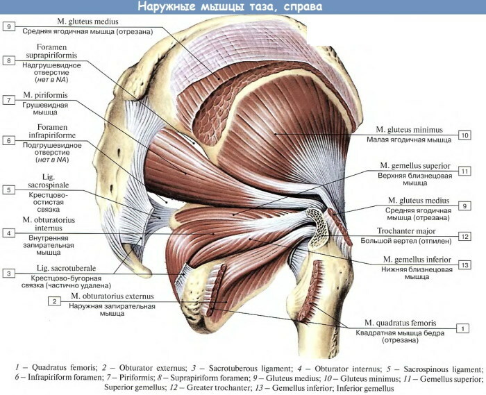
The anterior muscles of the zone adjoin the lower part of the large protrusion of the proximal epiphysis of the large femur. The group consists of the lumbosacral and pelvic muscles.
Posterior-outer group
Participates in the abduction of the hip joint. The posterior-external muscle group begins at the point where the lateral surface of the pelvic bone meets the iliac nerve process and the plane of the obturator membrane.
The latter is a thin plate of connective tissue that covers the anatomical opening of the skeletal structure. The posterior-external muscle group is attached to the round head of the femur.
The posterior-external group includes the following muscles:
- large gluteal;
- middle lumbar;
- small femoral;
- internal locking;
- fascia tensioner;
- pear-shaped;
- upper and lower twin;
- external lock.
The sacral plane, the fossa of the circular muscular head and the femoral epicondyle, belonging to this group, are considered the most vulnerable structural elements of the lower limb. They are most susceptible to injury. The posterior-outer group is responsible for tilting in the lower back, stabilizing the body in an upright position and maintaining an anatomically correct constitution.
Thigh muscles
Coordination of the knee and hip joints is performed. The muscle groups of the thigh have the largest specific gravity in the lower limb, significant length and significant strength indicators.
Such muscles are most easily amenable to development and hypertrophy by sports exercises. The thigh muscles perceive most of the load when walking, lifting weights, physical efforts.
A large artery passing in this zone feeds the entire leg, and channels conducting electrochemical impulses innervate the limb to the very fingers. The vessels departing from the muscle group prevent stagnation of lymph and blood, and ensure the functioning of numerous articular structures.
Front group
Consists of tailor muscle and quadriceps. The elastic fibers of the anterior thigh group are connected to the upper part of the plane of the iliac skeletal formation, and the lower part to the knee joint at the point of junction of the tibia muscle.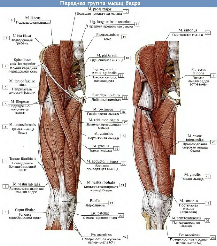
The sartorius muscle is responsible for flexion of both large joints of the limb. It rotates the lower leg inward and the femoral joint outward. The quadriceps muscle is responsible for leg strength, knee flexion.
The muscle of the human leg, called the quadriceps, occupies the entire area of the front surface of the femoral zone. In the photo with a description, its partial spread to the lateral plane is noticeable, the main functional features of the largest muscle of the lower limb are outlined.
The key anatomical significance of such muscles is to pull the thigh to the body when performing sports exercises, rotate the joint, and impart kinetic energy to the body when jumping.
The muscles of the medial group are located above the ischium with the trunk innervating the zone. They pass close to the pubic skeleton and cover the anatomical obstruction.
Composition of the medial muscle group:
- comb, attached to the proximal surface of the femur and responsible for bending the leg at a given angle relative to the body;
- thin, adjacent to the underside of the pubic symphysis (transitional connection between bone formations) and involved in the motor function of the knee;
- long adductor, starting from the medial plane of the femur and playing an auxiliary role in the rotation of the joint;
- short adductor, extending from the lower branch of the pubic segment of the skeleton and participating in the rotation of the limb;
- large adductor - attached to the ischial tubercle.
The medial muscles run along the comb line of the articular structure of the thigh. The main functional tasks are the abduction of the joint in the posterior plane and rotation in the same direction.
Posterior muscle group
Consists of semitendinosus, semimembranosus and popliteal muscles. It is customary to refer to this group as an independently functioning biceps muscle, which has significant strength capabilities.
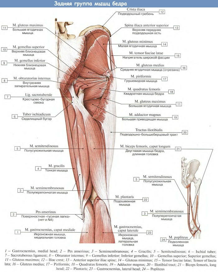
The musculature of such a thigh surface extends from the tubercle of the ischial zone. It extends to the lesser and tibial bone structures. Near the lateral edge, it forms a flaky structure, the surface of which is the gluteus muscles.
The muscle complex is attached to the tibial areas of the skeletal structure by tendon fibers. The functional purpose of such muscles is to provide mobility of the hip joint and ankle.
The tibial plexuses are responsible for the innervation of the muscular group. It feeds the musculature of the medial arterial bed, which bends around the femur. When flexing, representatives of the posterior group rotate the joint inward.
Calf muscles
The section of the lower limb contains the interosseous membrane, composed of cartilaginous and connective tissues. Anatomical design extends to the heel. The lower leg muscles are less developed than the thigh muscles.
Special sports complexes allow you to strengthen the muscles. The lower leg contains many small muscles and several large ones. They are responsible for the mobility of all joints of the leg, except for the hip.
A large number of tendon fibers and connective tissue structures are located in the lower leg area. The innervation parameters and blood circulation are similar to the muscles of the above-located zone, since it serves as their anatomical and physiological continuation.
Front group
Such muscles play a leading role in ensuring the mobility of the foot and turning it in different directions. The anterior group is responsible for abducting the anatomical support structure with the metatarsal bone in the vertical plane.
The muscles fix the ankle on the surface of the solid talus formation. The middle part of the head of the anterior group, together with the lateral plane of the femoral joint, maintains the tendon fibers in the anatomically correct position.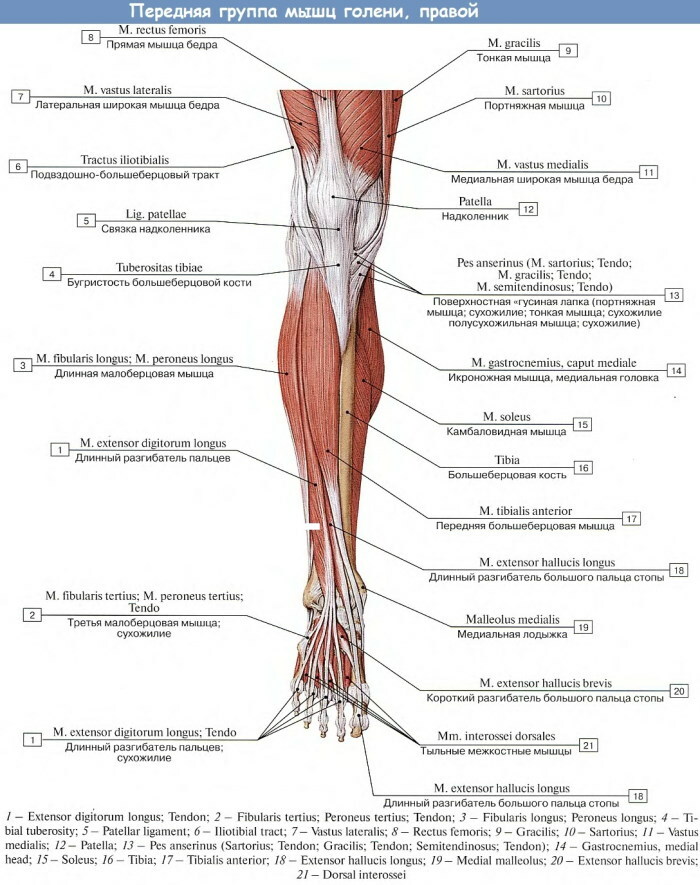
The muscles of the human leg of the photo with the description are presented in the form of intertwined elastic fabrics, often overlapping. The anterior muscular group of the lower leg has a similar structure.
It includes the tibial muscle and the long extensors of the toes. They supinate and pronate the foot, raising its medial edge. Different muscles are responsible for the mobility of the 4 anatomical processes of the limb and thumb.
Lateral group
The smallest of all muscle complexes of the leg. The lateral group is represented by 2 elastic fibers - long and short peroneal. The first muscle consists of a pair of heads extending from the bone of the same name to the condyle.
In the lower part, it smoothly passes into the connective-tendon tissue, enveloping the lateral part of the ankle in the posterior plane. Crossing the foot diagonally, the long muscle fits into the anatomical depression.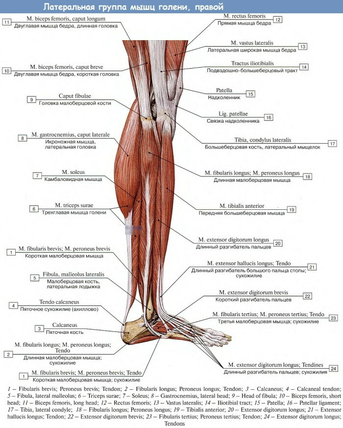
The muscle is attached by both heads to the protrusions of the bases of solid formations. Together with the short muscle, the long muscle performs pronation and supination of the supporting anatomical structure by raising its lateral edge and lowering the central axis.
Back group
The muscle complex consists of superficial and deep elastic fibers. The first are located near the subcutaneous fat layer. Such muscles of the posterior group are evolutionarily developed better than the deep-lying muscles.
The surface fibers give the calves their characteristic convex shape. The muscles of the layer stretch the capsule structure of the ankle joint. The back muscles of the lower leg extend from the plane of the femoral part of the skeleton and the upper segment of the iliac zone.
The group includes triceps and plantar muscles. The three-headed structure is a separate muscular complex formed by the superficial gastrocnemius elastic tissue and the deep soleus.
2 rounded heads depart from the first, and one from the second. Each of them has its own points of attachment to the osteoarticular capsule. In the lower part, the triceps of the lower leg forms the Achilles tendon and attaches to the tuberous protrusion of the hard heel structure.
The soleus muscle is responsible for stabilizing the body in an upright position and prevents it from overturning. It is most actively used when weighed down with a load. The calf muscle flexes the knee joint when the ankle is fixed.
Muscles of the foot
The complex consists of the smallest muscles. The fibers are transverse to the rest of the muscles. Such musculature is densely entangled with nerve tissues and blood vessels. This increases the sensitivity and makes the work of the support structure aimed.
The muscles of the human leg (a photo describing the complex demonstrates a large volume of fascia) in the foot area have a scant layer of subcutaneous fat. Elastic fibers have a complex pattern of physiological interaction with the small metatarsal bones.
Rear group
The ligamentous apparatus of the muscular complex constitutes the arch of the foot. The dorsal group includes the short extensors of the fingers. The flattened muscle originates from the superior and lateral planes of the anterior metatarsal bone.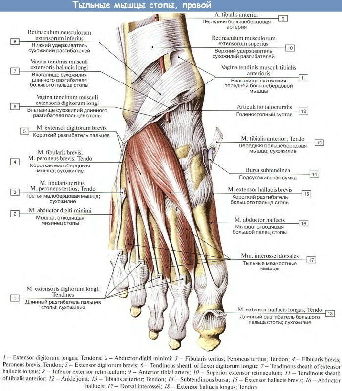
The short extensor and flexor muscles of the fingers transforms into 4 tendon processes as it moves. They are anchored at the base of the proximal, median, and distal phalanges. The muscle is woven into the fascia capsule of the dorsum of the foot.
A separate function is performed by the short extensor of the thumb, which is part of this complex. It lies in the individual anatomical bed, which prevents the migration of purulent exudate in the event of an inflammatory-septic process. The functions and structure of the muscle are similar to the previous one.
Has thinned and shortened tendon fibers. The muscular complex is fixed on the outer plane of the phalangeal bones. Medial elastic tissue is responsible for the mobility of the thumb. In addition to the shortened flexor, the group includes the adductor and abductor muscles.
The muscular complex is localized on the plantar surface of the support structure. The flexor short is located below the abductor muscle. It extends from the closure of the lateral longitudinal plane of the sphenoid bone with the plantar surface and a solid scaphoid formation.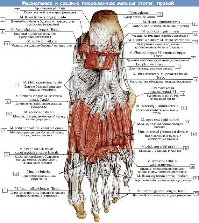
In addition to ensuring the mobility of the big toe, it is involved in giving stability to the arch of the foot. The adductor muscle is located under it. It stretches directly over the surface of the metatarsal bone. The muscle contains 2 heads - transverse and oblique.
Lateral group
For the innervation of the complex, there are bundles of sensory fibers located in the plantar part of the foot. The blood supply to the zone is carried out by the artery of the same name. The lateral muscle group is facing the front of the calcaneal tubercle.
The complex consists of only 2 muscles - the abductor 5th finger and the shortened flexor of the little finger. The first is responsible for the mobility of the central phalanx in 2 directions - lateral and along the longitudinal axis. The second muscle has a similar function for the little finger.
Middle group
Representatives of the complex supinate the anatomical support structure, attract the distal and lateral edges of the phalanges to it.
The composition includes the following muscles:
- flexor short, responsible for forward / backward movement of the fingers;
- square-plantar, which determines the longitudinal direction of the elongated flexor with tendons obliquely brought to the target fingers;
- vermiform, bending the central phalanges and straightening the nail;
- plantar-interosseous, responsible for the mobility of the fingers in the sagittal plane;
- dorsal-interosseous, abducting phalanges in the medial and lateral directions.
The last muscle is a separate complex formed by 4 elastic fibers. A photo of the middle group of a person's foot with a detailed description shows the structure of the dorsal-interosseous muscle. Each part of the complex performs an individual function of ensuring the mobility of the toes in all directions.
Videos about leg muscles
Leg muscles. Anatomy: