The human brain is a system that needs a fluid circulation, which performs many functions. This fluid is called cerebrospinal liquor, it is produced by special vascular plexuses of the lateral ventricles.
Total in a person in the central nervous system is not more than 150 - 160 ml of this fluid, which is incomparably less than the volume of circulating blood.
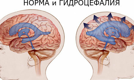
However, during the day, about 0.5 - 0.6 liters of CSF is produced, which should be absorbed near the venous sinuses of the hard shell of the brain in the pachyon granulations. In the event that there is a violation of the balance between the development of liquor and its absorption, hydrocephalus develops( see photo).
Contents
- 1 Brain hydrocephalus, what is it?
- 2 Causes of hydrocephalus
- 3 Internal and external brain hydrocephalus
- 4 Symptoms and signs of hydrocephalus in adults
- 5 Diagnosis of hydrocephalus
- 6 Treatment of hydrocephalus
- 6.1 Prognosis and complications
Hydrocephalus of the brain, what is it?
What is it?- Modern medicine defines hydrocephalus as an independent disease, or a complication in which the cerebrospinal fluid accumulates in the brain, as a result of which its movement through the cerebral and spinal cord pathways is disturbed.
Despite the relative simplicity of a formal definition, hydrocephalus can develop in many different ways. Therefore, neurosurgeons distinguish the following types of this pathology:
- Occlusal hydrocephalus. There is an obstacle in the course of the flow of the cerebrospinal fluid - occlusion. This can be a spike or, for example, a tumor. At the same time, rates of development and absorption of cerebrospinal fluid can be normal;
- Arerezive and disresorbative forms. In this case, the absorption of cerebrospinal fluid( resorption) is disturbed, as a result of which it accumulates;
- Hyper-secretory form. In this form, there is an excess of cerebrospinal fluid production, and the absorption "lags behind" in volume. As a result, the cerebrospinal fluid accumulates.
Convenient classification for doctors for the duration of the disease:
- acute hydrocephalus. The entire process from the first symptoms to gross cerebral infringements lasts no more than 3 days;
- subacute form - lasting up to 30 days;
- chronic - lasting from 3 weeks to 6 months or more.
In addition, hydrocephalus is also classified according to the level of liquor pressure. The process can be both normotensive and hypotensive.
Occlusal forms of acute hydrocephalus are often hypertensive, that is, they flow with a sharp increase in the pressure of the cerebrospinal fluid.
It should be noted that all the speculations about "increasing intracranial pressure" are meaningless if there are no clear signs of this process.
The only direct way to measure this pressure was and still is putting the manometer into the ventricles of the brain. Of course, for this you need to drill the skull bones.
This method is used for surgical interventions and shunting operations, which will be discussed below, but in outpatient practice they are used indirectly.
Causes of hydrocephalus formation
There are many reasons for the occurrence of hydrocephalus, but the main factors leading to this diagnosis - or compression by any volumetric formation, edema, or inflammatory and adhesive process. Most often, the following diseases and conditions lead to hydrocephalus, both in adults and children:
- Strokes. Compression of the cerebrospinal fluid can be either the volume of blood( intracerebral hematoma) or edema due to the ischemic focus;
malignant and benign brain tumors. Most often they are located inside the ventricles, next to the brain stem, or inside the trunk; - Infections of the central nervous system. Most often, these are purulent meningitis or encephalitis, tuberculosis, and other infections. As a rule, serous meningitis causes hypertensive hydrocephalus, which is well treatable;
- Central nervous system trauma: brain contusion, diffuse axonal injury, aneurysm ruptures, subarachnoid and subdural hemorrhages;
- Also, various metabolic and toxic encephalopathies( hypertensive, alcoholic) can be attributed to the causes of the development of chronic hydrocephalus.
Internal and external hydrocephalus of the brain
This formulation can be heard or read in the interpretation of MRI findings. Internal hydrocephalus is determined by the increase in the ventricles and median unpaired cerebrospinal fluidways, external hydrocephalus refers to the "circumference" and peripheral space.
Mixed cerebral hydrocephalus often develops. It is important that these forms are open, with all liquor pathways being passable, and the fluid flow is not disturbed.
If there is an obstacle, as with occlusion, then a closed form of the disease develops.
There is also a substitute hydrocephalus of the brain in adults, at which the gray matter of the brain( that is, the bark) is replaced by a cerebrospinal fluid that circulates in the subarachnoid space of the convective surface of the cerebral hemispheres.
This occurs as a result of atrophy of the cortex, and not due to hydrocephalus, that is, atrophy is the primary process. Therefore, this term is gradually becoming obsolete.
Symptoms and signs of hydrocephalus in adults

In neurology, in addition to focal symptoms, in which the function of any department of the nervous system suffers, there is a general cerebral symptomatology that is present in hydrocephalus.
So, for example, in the acute form of of rapidly developing occlusive hydrocephalus( for example, with tumor dislocation, or in case of a clotting with purulent meningitis) the following symptoms will be expressed:
- Intolerable, "bursting" headache, on all sides, without a clear localization. The patient feels that he is "pumped up his head with a pump."At the same time, headache intensification in the morning hours and relief of feeling for the evening, or in the afternoon, are characteristic.
- The appearance of nausea and vomiting, in more severe cases with the development of hypertensive forms, can be "cerebral vomiting" - vomiting "fountain" without any previous nausea, which becomes completely unexpected for the patient. This is due to irritation of the receptors of the bottom of the 4 ventricles, or "diamond-shaped fovea".This vomiting does not bring relief;
- When examining the fundus, stagnant discs of the optic nerves are noted;
- In case of deterioration, drowsiness, sopor and coma occur, which can result in dislocation and edema of the brain substance. This process is dangerous with different variants of wedging of the vital ancient centers of the brain, which are localized in the trunk and the medulla oblongata, and are responsible for respiration and circulation. A classic example is the insertion of the tonsils of the cerebellum into the large occipital opening. This leads to death.
For chronic hydrocephalus , which lasts for many months against the background of organic damage to the central nervous system, there will be very different symptoms:
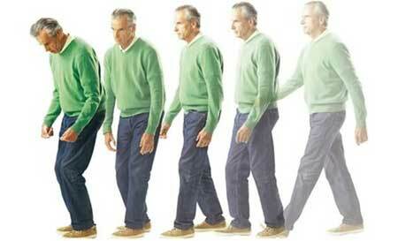
- The manifestation of progressive dementia;Instability of gait, or leg paresis( the so-called peripheral lower paraparesis);
- Urination disorders.
This picture can be observed with various senile disorders on the basis of chronic intoxications, for example, in deep alcoholics "with experience".
In this case, the development of the chronic form does not manifest sharply, but develops gradually, often the first symptoms appear one month after the transferred disease, for example, a stroke.
Patients wander wakeful and sleep patterns, initiative and activity decrease, they become listless and indifferent, memory and attention begin to deteriorate, then gait disturbances appear, and in the ending, urinary incontinence joins, and in severe cases, feces.
Diagnosis of hydrocephalus
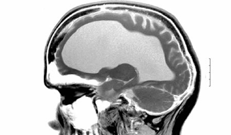
Currently, the diagnosis is not particularly difficult. Visualizing techniques( CT, MRI) allow us to draw conclusions on indirect grounds. So, according to the MRI of the brain, hydrocephalus can be exposed to the expanded lateral ventricles.
Nevertheless, each of these methods has its own "advantages": a computer X-ray tomography reveals the exact contours and boundaries of the cerebrospinal fluidways, allows you to accurately calculate the size of the ventricles. Magnetic resonance imaging by the reaction of the brain tissue allows us to clarify the degree of severity of the process.
Of course, the study of the fundus, echoencephalography and direct measurement of cerebrospinal pressure, which is performed during neurosurgical operations, remains important.
Treatment of hydrocephalus of the brain
As in many other cases, there is conservative and surgical treatment. External hydrocephalus of the brain in adults, treated like the internal one, implies the following therapeutic tactics:
- Use of osmotically active diuretics( urea, mannitol in infusions);
- Diacarb is a diuretic drug that inhibits carbonic anhydrase and is used specifically to increase intracranial pressure in outpatient practice;
- Inflammation of glucocorticosteroids, for example, dexamethasone, is helpful in the inflammatory response( for example, in meningitis).Analgesics are introduced symptomatically, as well as barbiturates, which protect brain tissue from hypoxia, reducing its need for oxygen.
In case the conservative treatment of hydrocephalus of the brain was unsuccessful, surgery may be required.
So, with acute hydrocephalus, it is required to "adjust the outflow", which will relieve the ventricular system and reduce the pressure. For this purpose, external ventricular drainage is performed.
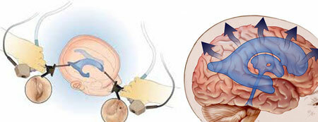
Shunting of the brain in hydrocephalus is a method of treating chronic hydrocephalus, in which excess cerebrospinal fluid is "dumped" where it does not interfere and is quietly absorbed. For this purpose, a catheter is installed in the ventricles of the brain, which is connected to a valve that allows "bleeding" the excess of CSF when certain pressure values are reached.
After its lowering, the valve closes. Further, a long catheter is attached, allowing the withdrawal of the cerebrospinal fluid, for example, into the abdominal cavity, where it is absorbed. This type of bypass is called ventriculoperitoneal.
Prognosis and complications of
Before performing a bypass operation, it is necessary to check whether the patient causes an improvement in the reduction of the CSF volume. To do this, perform a lumbar puncture and take about 40 ml of CSF.In the event that the patient is feeling better, it makes sense to do a bypass operation. If there is no effect, then other ways of treatment should be sought.
The most dangerous complication of hydrocephalus, which was mentioned above, is the development of edema - swelling of the brain and the dislocation of the median structures. The symptoms that indicate the onset of this formidable complication are a gradual loss of consciousness, starting with drowsiness, as well as dilating the pupil on one side after a short-term constriction, convulsions, fever( hyperthermia), and pyramidal insufficiency.
Therefore, such patients should urgently be hospitalized in the department of neurosurgery.
In chronic hydrocephalus, the prognosis for life is favorable with appropriate timely treatment.