Women are at risk for developing nephroptosis - the prevalence of this condition is 1, 5%.In men, it is diagnosed only in 0.1% of cases.
Right-sided omission often develops. Nephroptosis of the left kidney, as well as a bilateral anomalous position, is very rare.
Contents of
- 1 Kidney nephroptosis - what is it?
- 2 Kidney nephroptosis causes
- 3 Nephroptosis stages
- 4 Kidney nephroptosis symptoms in stages
- 5 Treatment of kidney nephroptosis
- 5.1 Disease of the disease
- 5.2 Forecast
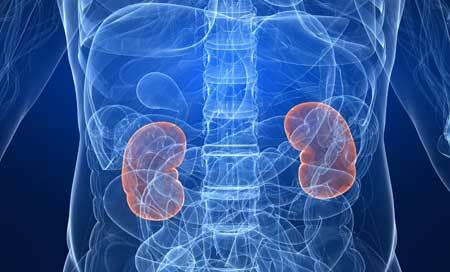
Nephroptosis of the kidney - what is it?
In a normal state, almost all organs of the human body can be displaced relative to a typical anatomical location. But if such changes exceed the permissible limits( norms) and begin to interfere with the functioning of the organ - the displacement is considered pathological.
Similarly, the kidneys can move within the normal range, with the movement and movement of the body. Such mobility favorably affects the urinary process.
But in some cases this movement may become abnormal( in medicine it is called nephroptosis).
Among all kidney diseases, nephroptosis is a fairly common but poorly diagnosed condition. In translation from Latin "ptosis" means omission, "sliding down".In reality, any pathological change in the position of the kidneys, and not just the true omission, is called nephroptosis.
The kidney can "wander", that is, move every time to a different place, can go down, reaching the small pelvis, can change the position around its axis.
Most often, nephroptosis of the right kidney is due to the initially lower location of the kidney due to the fact that the liver is located above it. In addition, the ligaments holding the organ on the right side are weaker than the left.
Causes of kidney nephroptosis
The structures that affect kidney retention in a typical anatomical location are ligaments, fatty tissue and abdominal wall muscles.
Therefore, the factors that lead to the appearance of nephroptosis of the right kidney will be all those factors that contribute to the weakening and alteration of restraining structures, as well as some anatomical features. The latter include:
- Underdevelopment or absence of lower ribs;
- Congenital malformation of the kidney location associated with embryonic developmental disorders;
- Asthenic( lean with long stature) type of build;
- The change in the proportions of the body, observed during the period of intensive growth of the body( as a rule, it is a pubertal with the intensive hormonal changes typical for it).
Reducing the amount of adipose tissue is another important reason for the acquired nephroptosis. This can occur with intensive weight loss within the framework of an improper diet program or after an exhausting infectious disease.
Injuries to the ligamentous apparatus are a direct weakening factor of the restraint system. They appear because of a sudden change in intra-abdominal pressure or a change in the position of the body. This predisposes such sports as basketball, football, volleyball and bodybuilding.
Therefore, people at risk for nephroptosis, as well as having the congenital form of this disease, these physical exercises are contraindicated. They further exacerbate the improper position of the kidneys. Trauma of ligaments can also be observed when falling from a height, striking the lower back or abdomen. In this case, the ligament ruptures with the formation of extensive hemorrhages.
Many women during pregnancy can develop a nephroptosis on the right. This is facilitated by several predisposing factors, the dominant role of which in different trimesters varies:
- A sharp drop in intra-abdominal pressure after delivery;
- Large abdomen during pregnancy;
- A large number of pregnancies and childbirths in the anamnesis;
- Hormonal rearrangements( among them, especially significant increase in estrogen levels).
Representatives of some professions have an increased risk of developing kidney ovulation throughout life. These are the specialties that are associated with:
- Vibration and shaking( drivers);Excessive physical strain( loaders);
- Long standing( hairdressers, surgeons).
Stages of nephroptosis
The stages of the disease reflect the range of the kidney displacement relative to its normal position, the presence of changes in its structure, the functional state and severity of existing complications.
The first stage of is characterized by a small displacement of the kidney in the vertical position of the person. Determine the nephroptosis of the 1st degree of the right kidney visually does not work. In thin people, you can feel the displaced kidney through the anterior abdominal wall in a standing position in the period of maximum inspiration. Functional kidney disorders in the first stage are absent.
In the second stage of , the kidney is visually determined from the right under the ribs in the standing position. However, when the position of the body changes from vertical to horizontal, it leaves the field of vision, hiding in the hypochondrium. At palpation the doctor can easily fix it there.
Because of the greater displacement of the kidney in the second stage, its rotation relative to the longitudinal axis is possible. This leads to an inflection of blood vessels and ureter.
As a result, the flow of arterial blood is disturbed with the development of oxygen starvation( kidney ischemia).Along with these, the venous outflow also suffers, leading to increased pressure in the renal vein. Violation of the outflow of urine, caused by the excess of the ureter, promotes the development of infection. Therefore, often enough at this stage, chronic pyelonephritis is attached.
In the third stage of the , the kidney, regardless of the position of the body and the breath-exhalation phase, is in the right hypochondrium. When the disease progresses, it descends into the small pelvis. Virtually all patients with stage III nephroptosis develop chronic pyelonephritis. Vascular and metabolic disturbances in the renal parenchyma are also aggravated.
Symptoms of kidney nephroptosis in stages

In the first stage of kidney nephroptosis, symptoms may be absent or episodic, and their severity is minimal. The pain that appears in the lower back does not have specific signs. It is dull or aching, it does not stand out in anything.
Therefore, the first stage is often left unattended by the patient, or its symptoms are written off for manifestations of osteochondrosis, myalgia or chronic adnexitis in women( inflammation of the ovaries).
People with a risk of developing nephroptosis need to pay attention to one important feature. It consists in the fact that pain occurs when the position of the body changes - it appears and becomes stronger standing, and lying alone is stopped without the use of medication.
A similar situation is observed with physical activity - with pain there is pain, but at rest - no. The further the kidney begins to "walk", the lower the pain senses descend. So, over time, the pain moves to the sacrum, the lower part of the abdomen. Simultaneously, it becomes more intense, and episodes of it are repeated more often.
In the second stage of , pain appears even with little physical exertion, for example, when climbing a ladder. Disturbance of blood supply and stagnation of urine, characteristic of the second stage of nephroptosis, lead to the development of complications, as well as the appearance of protein and erythrocytes in the urine.
The adherence of pyelonephritis contributes to aggravation of pain, weakness, fatigue, apathy and fever.
The third stage of nephroptosis has all the symptoms of a developed disease with complications. The pain becomes permanent, poorly controlled by analgesics. This leads to psychological depression of the patient. Against this background, depression and neurasthenia may occur.
Problems with digestive system, manifested by vomiting and diarrhea, decreased appetite. Physical load becomes dangerous, becauseshe threatens with the appearance of renal colic, associated with a significant kink of the ureter. At this stage, clinical manifestations of chronic pyelonephritis( pain, changes in the analysis of urine), which become even more pronounced, persist.
Constant increase in pressure in the renal vein, caused by its compression, leads to the rupture of small vessels and the appearance of blood in the urine.
The inflection and narrowing of the renal artery activate the juxtaglomerular complex that produces angiotensin. This leads to an increase in total blood pressure and related complications. At this stage, hydronephrosis sometimes develops.
The final diagnosis of nephroptosis of the right kidney is based on a thorough survey, identifying the nature of the pain, time and conditions of their occurrence. Palpation( probing) of the region of the right hypochondrium and the determination of the displaced kidney indicates the stage of the process.
Laboratory methods include:
- General blood and urine analysis
- Biochemical blood test with determination of urea, creatinine and total protein concentration. These indicators help to judge the safety of kidney function. In some cases, a sample of Reberg is required - an estimate of the rate at which the kidneys filter blood per unit time.
Instrumental studies are also shown to patients with nephroptosis: the
- kidney ultrasound in a vertical position allows you to visually assess the displacement;
- X-ray examination with contrast( urography) is the main method of nephroptosis diagnosis,allows directly to see the omission and determine its degree;
- Angiography of renal veins and arteries allows you to clarify the state of the vessels and the position of the kidney;
- A radioisotope study helps to conduct a functional assessment of the kidney.
Treatment of kidney nephroptosis
After confirmation of the diagnosis of kidney nephroptosis, treatment depends on whether there are complications or not. If they are absent, then conservative therapy is indicated. It is aimed at strengthening the supporting apparatus of the kidney. For this purpose, the following are recommended:
- Wearing a medical bandage( dressed in the prone position before getting out of bed
- Strengthening the muscles of the anterior abdominal wall with the help of specially selected physical exercises
- High-caloric diet food to increase the percentage of fat tissue in case of severe depletion or significantasthenia
The development of complications raises the question of the need for surgical intervention, it is carried out according to the following indications:
- The constant nature of pain that can not be bought
- Changing the quality of life associated with the presence of pain syndrome
- Chronic pyelonephritis, which threatens the development of renal insufficiency;
- Significant deterioration in functioning of the displaced kidney;
- Persistent increase in blood pressure, requiring the use of antihypertensive drugs;
- Persistence of hematuria(the presence of blood in the urine), indicating an increase in pressure in the renal veins;
- Hydronephrosis is an expansion of the kidney associated with stagnation of urine.
Operative treatment consists in conducting nephropexy - fixing the kidney in its typical place with preservation of physiological mobility. At present, this operation is performed by the laparoscopic method, which is the least traumatic and cosmetically more beneficial for the patient.
Conducting conservative treatment is aimed at changing the way of life in order to stop the progression of the disease. Medical measures should be carried out constantly along with preventive examinations at the urologist. Every year, patients with nephroptosis are shown an ultrasound examination, even after surgical correction, since there is always a risk of repeated omission.
Danger of disease
The displacement of the kidney is dangerous in view of the possible development of disorders from the renal vessels or ureter. Twisting and squeezing of the arteries and veins leads to oxygen starvation of the kidney tissue, increased venous pressure. And this, in turn, provokes a trauma of small vessels inside the kidney, further exacerbating the existing disorders.
The kink of the ureter threatens with a dangerous delay in urine and the development of inflammation of the kidney tissue. Prolonged and constant disturbance of microcirculation leads to metabolic disturbances in the renal parenchyma. So the vicious circle closes.
Prognosis for
The prognosis for patients with right-sided nephroptosis depends on the presence of complications and the degree of omission. The appearance of renal failure is considered a poor prognostic sign, becauserequires the patient's transfer to hemodialysis( artificial blood purification).
Therefore, the main task of the urologist is to diagnose the progression of the disease in time and take all measures to prevent it. This means that it is necessary to determine the indications for the surgical treatment of nephroptosis in a timely manner.