technique The valsalva test for varicocele is used by medical professionals around the world. This technique allows you to identify the presence of problems with the veins in the early stages. Also, this method is used to identify various problems with the cardiovascular system. With the help of a special method, doctors can establish negative changes in the absence of visible symptoms. For this reason, experts recommend that you immediately visit a medical center for intimate problems.

Characteristics of an inguinal circulation
In order to understand what a sample is for, it is necessary to understand the characteristics of varicocele. This pathology affects the venous bundle, which carries the blood fluid to the testicles. The accumulation of veins forms a tangle that resembles a bunch of grapes. Due to this feature, the tangle is called the lobate plexus. These fibers are responsible for the correct activity of the male sex glands.
Blood flows through the veins to the organs. In the blood fluid contains a large number of red bodies. These cells have a biconcave form. This structure of red blood cells is due to its function. In the indentations on the surface of the erythrocyte, an oxygen molecule is transferred. When the erythrocyte enters the tissue, the molecule leaves its surface. Oxygen passes into the cells of the organs. With a sufficient amount of this substance, the tissues are capable of self-renewal. The aged cells are replaced by young cysts.
Varicocele refers to a variety of varicose veins. Varicose veins are characterized by the formation of a stretched pocket on one of the sections of the vessel. Pathological process occurs under the influence of various causes. Under the influence of negative factors, the elasticity of the fiber becomes smaller. The blood lingers on this site. Gradual accumulation of blood leads to a strong stretching of this zone.
Because of the pocket, the blood circulation is reduced. The pressure on the affected area increases. The lack of oxygen leads to a disruption in the functioning of the organ fed. Lozovidnoe plexus carries oxygen to the paired sex glands. With a shortage of nutrients, spermatogenesis deteriorates.
The testicles are responsible for producing healthy germ cells. Spermatozoa should have a number of specific qualities that contribute to fertilization. With varicocele, spermatozoa lose their ability to move. This causes a massive death of germ cells. A man becomes barren. It is for this reason that it is important to determine the presence of varicocele in the early stages. If the disease turns into a severe form, there is a risk of death of one of the paired glands. Doctors distinguish three stages of the disease. The first stage of varicose veins is characterized by the formation of a densified section on the wall of the vessel. In this case, the patient has no visible symptomatology.
Anxiety in men causes the second stage of the disease. At this stage, the formation of the pocket. In this case, the patient has the first symptoms of the disease.
A dangerous form of the disease is the third stage. At this stage, the walls of the pocket become very thin. There is an increase in blood pressure. Damaged tissue may burst. Hemorrhage in the abdominal cavity can lead to death.
Signs of varicose disease

problem In order to determine the presence of a pathology in time, it is necessary to know its symptoms. At the first stage, many men do not pay attention. This leads to further aggravation of the situation. Doctors recommend paying attention to the following visible symptoms:
- soreness in the scrotal area;
- swelling of one of the testicles;
- cyanosis of the skin of the groin;
- erectile dysfunction.
The first visible sign of a pathology is soreness in the scrotal area. The appearance of pain increases with certain actions of the patient. Unpleasant sensations occur with prolonged walking or active physical activity. Also, soreness accompanies the process of ejaculation. A feature of this symptom is its complete disappearance when staying in a horizontal position.
With increasing intensity of the disease, edema of one side of the scrotum is observed. Often, puffiness appears on the left side. This localization is characterized by the peculiarities of locating the lobate plexus. In this zone, the venous bundle forms a right angle with the vas deferens. In rare cases varicocele appears on the right side of the scrotal sac.
Specialists also note that due to a decrease in blood flow to certain parts of the vein, the color of the skin changes. On this zone there is a cyanosis. Also, when the palpation of this area, the patient notes increased pressure in the abdominal cavity.
Pathological process is accompanied by various violations of sexual function. Lozovidnyj the ball feeds testicles and vas deferens. During ejaculation, the seminal fluid passes through the ducts. Against the background of a strong blood pressure, the volume of sperm released can decrease.
Also, problems are detected and with sexual arousal. Erection occurs when filling cavernous bodies with blood. The fluid is fixed in the cavernous bodies with special sphincters. With insufficient blood volume, the erection becomes inferior. Also, spontaneous removal of blood from the penis during sexual contact can occur.
If a man has noticed one of the listed signs, he needs to seek advice from a specialist. Only physicians can accurately determine the cause that caused these symptoms.
Causes of varicose veins

Often, patients are asked what causes the negative changes in the vascular system. The causes of pathology are diverse. The following negative factors are considered, entailing the expansion of the vessels of the inguinal zone:
- genetic predisposition;
- active physical activity;
- professional features;
- is an inflammation of the pelvic organs.
It was found that varicose veins are found in patients because of genetic predisposition. If older family members suffer from this disease, the younger generation can also acquire this problem. In this case, doctors advise to take preventive measures. Only prevention can reduce the likelihood of developing varicocele.
Men who are active in physical activity are at risk. At high physical loads, the pressure in the vessels increases. After the end of the exercises, it will return to normal. Frequent expansion and narrowing of the lumen of the fiber is fraught with stretching of the tissue. This entails the formation of a stretched portion of the vascular wall. To reduce the risk of developing health problems, all exercises should be performed only under the supervision of a sports coach.
Affects the blood circulation and the patient's profession. In many people, activity is accompanied by a long stay in one position. This posture is accompanied by a decrease in the intensity of the circulation. The liquid rotates more slowly. There is stagnation in the small pelvis. This causes a gradual expansion of the venous bundle.
Diagnostic features of
When a pathology is detected, a specialist assigns diagnostics. You can identify varicocele in several ways. Dopplerography is a common diagnosis. This study allows you to establish the strength of the passage of blood in different parts of the vessels. With dopplerography on the screen, you can set the patency of veins with a special solution.
Electroencephalography is also used. This technique is carried out with the help of a special apparatus. Electric waves are passed through separate zones. When a wave collides with an obstacle, a pattern appears on the screen. Varicocele is characterized by the formation of one or more nodes on a vesicular beam.
Features of the
methodA more accurate method is a test of valsalva. This test was previously used to determine the functioning of the middle ear. Also, the technique helped the body prepare for immersion to a greater depth. Divers using this method avoided a sharp drop in blood pressure.
Has found a sample and application in the phlebological field. It was found that with a delay in breathing, a certain change in the activity of venous sphincters occurs. With a deep breath, the fluid is fixed in the veins. The walls are easily palpated. This feature is typical for the vertical position. In the horizontal position, a sharp outflow of blood takes place. The pressure decreases. Both properties allow specialists to carefully study the state of vascular fiber.
The valsalva test was known in the 19th century. Developed by her Italian scientist. Antonio Mario studied the work of the hearing aid. He noticed that the membrane retains its shape at the same pressure in the external environment and the auditory tube. The pipe performs the function of a kind of vent. It helps the air to pass into the middle ear to maintain constant pressure in it. In this case, the pressure drop is observed with swallowing movement. The growth of external pressure on the membrane is observed when moving to a greater depth. To reduce the negative impact on the membrane and avoid its rupture, scuba divers forcibly exhale. This helps to normalize the pressure on both sides of the membrane.
Valsalva found application of this test in phlebology. With a deep breath, the sphincter of veins is closed. Blood is fixed in the vessel. This allows us to study the maximum diameter of the venous lumen. During exhalation, the total volume of fluid and exhaled air is examined. With the help of a valsalva test, the presence of pathological sites can be accurately determined.
The sample is effective in varicose veins of peripheral organs. It is also used to study the operation of the respiratory system and the hearing aid.
The course of the study
The varicocele is a vascular response when inhaled. When the air is delayed, there is a strong tension in the peritoneal wall. When the tension of the wall of the plexus plexus is bulging. A specialist can palpate the beam along its entire length. When the wall leaves, they fall off. This helps the doctor to detect even small nodular formations.
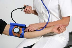
A special tube connected to the pressure gauge is used to conduct the Valsalva test. Before starting the procedure, the patient must take a horizontal position. In a lying pose, the man inhales deeply. There is a fall in the venous bunch. The doctor performs palpation. After that, the patient takes the tube and tightly compresses her lips. At the same time, a gradual exhalation takes place. The device captures the pressure of the output stream. In a healthy person, the maximum value should not exceed 40 U / min. If the indications are higher, there is a pathology.
After the procedure, the patient assumes an upright position. All manipulations are repeated in the same order. According to the results, the doctor can draw certain conclusions.
Not all specialists can accurately determine the presence of changes in the venous bundle. To confirm the results, the sample must be accompanied by one of the additional methods. More accurate results are obtained by the joint implementation of the sample with Doppler. If a seal is found on one of the sections of the vessel, the doctor applies Doppler. It helps to accurately determine the size of nodule formations and their localization.
Palpation is also necessary. The palpation is carried out over the testicular region. When palpating, you need to determine the condition of the vas deferens and the plexus plexus. On the inhalation, the scrotal of the affected testicle is carefully examined. The walls of the ducts must be smooth and tense. If this is not present, the ducts receive insufficient amounts of blood fluid.
Advantages of the
method Many modern specialists in varicocele research prefer the method of valsalva. This phenomenon arises from the presence of several advantages:
- ease of holding;
- obtaining a reliable result;
- early diagnosis of the problem.
No additional instruments are required for the sample. The patient also does not have to follow any rules. The whole exercise is carried out with the help of one small device.
When the vascular fiber is protruded, the physician can carefully study its structure. The presence of a small lesion or densification is quickly revealed. Also, with palpation, it is possible to detect changes in the activity of the vas deferens.
With the help of a test of valsalva, any stage of varicocele can be detected. Negative changes are diagnosed in the early stages of the disease. This gives an advantage over other diagnostic methods.
Any pathological disturbances in the functioning of the vascular system affect the activity of the organs fed. In varicocele, negative changes occur in the reproductive system. Timely detection of the problem can quickly stop it. For this reason it is important to undergo a preventive checkup every six months.