 The concept of bone cancer combines all types of human tumors that affect the bones of the skeleton.
Several varieties of benign growths are distinguished here, which can subsequently develop into the malignant stage, as well as malignant formations required for immediate removal and long-term treatment.
Among all benign tumors, osteoma is more common.
Content of the article
- Concept and statistical data
- Causes and risk factors
- Possible localization of education
- What types of tumors are there?
- Knowledge of the symptoms is the key to successful treatment
- Diagnosis of
- To treat or not to treat is the question
- Prognosis is favorable
- Necessary prevention measures
Concept and statistical data
Osteoma is a benign tumor that is often exposed to children and young people under 20years.
The presented disease rarely turns into a malignant form. It consists of cells of bone tissue. Characterized by a slow flow, does not entail the formation of metastases or germination in the surrounding soft tissues and organs.
The appearance of a tumor for a long time may not show signs of existence. An exception may be intracranial growths, which in the process of growth and development squeeze the brain, which leads to severe headaches.
Localization in other parts of the body leads to a cosmetic defect and the patient's treatment to the doctor.
Causes and risk factors
In half of cases, osteoma occurs in children due to genetic "transmission".
If a child's parents suffer from such a disease, they must take measures to prevent the occurrence of a tumor in their child.
Due to the slow formation and development of the tumor, experts recommend that the examination be performed every year.
In addition to the genetic predisposition, the following causes of the presented ailment are distinguished:
- congenital predisposition of - a newborn can have superficial bone formation;
- the presence of any diagnosed connective tissue diseases , for example, rheumatic manifestations;
- is the diagnosed gout , which has causes of metabolic disorder;
- the presence of an infectious disease in a person ;
- transferred bone injuries .
Potential patients should be cautious if they have a diagnosis of the problems described above.
Possible localization of education
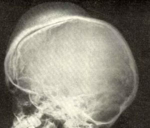
In most cases, single formations on the head are identified.
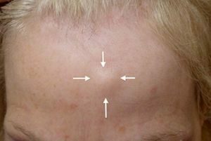
On the photo of the osteoma of the frontal bone
Osteoma of the frontal bone is often diagnosed - according to statistics it is about 52% of the diagnosed cases, 22% of the detected tumors are located in the frontal sinus, namely in the trellis labyrinth.
In the maxillary sinuses, osteomas are observed in 5% of cases. In the rest of the human body, this type of tumor is extremely rare, with a large proportion of tumors with localization inside the skull.
Also specialists give the following statistics, according to which the tumor on the forehead is manifested in men 2 times more often than in women.
At the same time, osteoma of the nasal sinus is diagnosed 3 times more often in women, rather than worried about men.
What types of tumors are there?
The osteoma is divided into three types:
- Solid - consists of dense concentric plates located parallel to the surface of the bone. Their density reaches the elephant bones.
- Spongy - consists of porous substances.
- Brain - most of all the substances that formed the build-up, is the bone marrow.
Also identified osteomas can be divided into two groups:
- Hyperplastic growths - are formed from bone tissue. In turn, exostoses( an outgrowth on the surface of the bone) and enostoses( a tumor inside the bone, which "bursts" from the inside) are isolated.
- Heteroplastic neoplasms - are formed in the tissues of internal organs or muscles. Later, their development is localized at the junction of the muscles to the tendons.
Accurate and high-quality diagnostics will help to identify tumor formation and determine its appearance, which significantly affects the further treatment.
Knowledge of symptoms is the key to successful treatment of
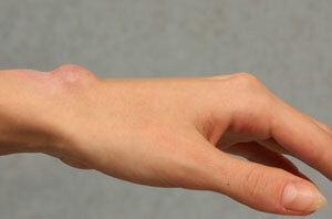
 Osteomas in the initial stages do not cause pain, so often the patient pays attention to a small tubercle - a seal on the forehead or in another part of the body that is characterized by a palpable palpation, but rather tight density.
Heteroplastic neoplasms lead to a pain syndrome, more similar to the symptoms of internal disease.
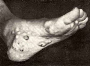
If the tumor has a beginning in the muscles, the person feels pain, which is often explained by simple physical exertion.
In the case of localization of education within the skull, a person may suffer:
- headaches;
- with seizures of epilepsy, which had not previously been diagnosed;
- absentmindedness, memory impairments with short-term loss.
The localization of education inside the nasal sinuses entails shortness of breath, which leads the person to the clinic for a survey.
Diagnosis of
The tumor is diagnosed with an external examination of the physician by palpation. 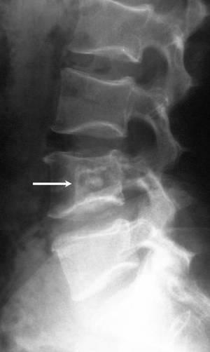
Confirmation must be submitted in the form of a completed X-ray examination.
In the picture, the doctor will be able to see the distinct manifestations of the tumor.
Additional diagnostic methods are often used:
- computed tomography allows accurate determination of tumor size and location;
- radioisotope scanning of the skeleton allows to determine the type of formation;
- MRI - is often used instead of X-ray examination if a heteroplastic lesion is observed.
After all tests, the doctor can accurately determine the nature of the tumor. For successful treatment it is necessary to know its further development in dynamics.
To treat or not to treat is the question
Treatment of osteoma does not always involve its removal.
If the tumor is localized in a "hard-to-reach" place, doctors decide not to touch the patient and only observe his condition and the further behavior of the build-up.
Here we consider such an aspect as the increase in education and the possible harm to the body and the general state of man.
The doctor should be consulted annually for the examination, as well as when the condition worsens, when it is noted:
- the patient began to feel pain during movement or palpation;
- there is a violation of joint mobility;
- the patient noted the presence of inflammation in the affected area.
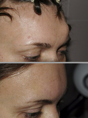
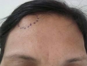
Before and after surgery
In such cases, oncology doctors decide to remove the tumor, wherever it is.
Because such signs often indicate malignancy of the tumor.
When the tumor is located on the outer surface of the bone, its removal has a cosmetic overtones, because the built-up edge can have significant dimensions and disfigure the appearance of the person.
The only method of treatment is the surgical removal of the tumor .
Along with the build-up, part of the affected bone is removed, which sometimes requires additional prosthetics by "attaching" the implant.
Prognosis - favorable
With the timely removal of the tumor itself and the partial resection of the periosteum and bone tissue, the recovery prognosis is more than favorable. In such cases, relapses rarely occur, which affects the positive treatment.
Removal of a tumor on the face and other visible places does not lead to a cosmetic defect. A small scar is the only reminder of a transplanted operation.
Necessary prevention measures
As far as prevention is concerned, people who fall into the risk group should be especially attentive to themselves.
If your family and friends suffer from the presented disease, go through a regular examination to identify tumors that for a long time will not show signs of their existence in the human body. The same actions should be carried out by people who have already undergone an operation to remove osteoma.
Bone cancer is not a verdict. Osteoma is an initially benign tumor. With a careful attitude to their health, a favorable outcome of events is quite possible.