 Ganglia with an armor."Knot" - in normal anatomy, a neural tissue containing neurons and their processes - axons and dendrites - is designated.
But according to the international classification of diseases of the world health organization, under the code "ganglion" in ICD-10, any degenerative-dystrophic disease of the wrist joint of the hands is concealed.
Also in the literature you can find the names "ganglion", "cyst", "swelling", "hygroma".All this is the name of the same pathology - the ganglion.
That is, it is a benign tumor-like process, which is formed due to the growth of synovial tissue. It differs from hygroma in smaller sizes.
In 80-90% of cases, all benign lesions of the hand are hygromes. Especially often this pathology affects young girls and women( frequency about 60%).
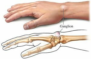
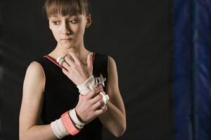
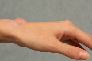
Externally, the hygroma of the hand represents a tumor-like formation on the palm of the hand or the back of the wrist. By consistency, it can be dense or more elastic. Stretched in length and, as a rule, painful.
Contents of the article
- What are the causes of the violation?
- Pathology development mechanism
- Species of formations
- Where can be located?
- Characteristic manifestations at different stages of the pathology
- Diagnosis
- How to get rid of the problem?
- Laser removal - effective innovation
- Possible complications
- Preventive measures
What are the causes of the violation?
Reasons that can lead to the development of education:
- permanent mechanical trauma of the same place;
- professional overload brush;
- hereditary predisposition - it is believed that in 25-30% of cases there is a family precedent;
- hemorrhage in the cavity of the articular bag;
- pressure difference inside and outside the joint;
- infection of the joint space, especially specific pathogens, for example, a tubercle bacillus, actinomycosis, syphilis, etc.
Pathology development mechanism
The joint capsule consists of a connective tissue. From the inside it is lined with fibrous litter.
Articular fluid enters the joint through valves from the joint space. This movement occurs in one direction, that is, only from the periarticular bag into the joint cavity. The outflow is not reversed. This liquid persists in the joint cavity and is partially processed by the cells of the same fibrous connective tissue.
After injury or exposure to another etiological factor, degenerative-dystrophic processes occur in the tissue, that is, the death of the cells that make up this tissue.
Therefore, it becomes impossible to process excess synovial fluid, it thickens, thickens and looks like jelly in consistency.
Over time, this fluid can build up the connective tissue, forming a cyst. Often densely attached to the underlying tissues. Over time, calcium capsules can be deposited on a capsule that delimits a defect. E
will lead to calcification, that is, if the consistency of the defect was previously elastic, now it becomes gradually firm.
Varieties of formations
By the number of chambers that are formed, these ganglions are distinguished:
- single-chamber;
- multi-chamber.
Where can it be located?
Localization may be affected:
- articular phalanx;
- wrist joints;
- wrist joints.
On the feet:
- joints of phalanges;
- joints of a metatarsus;
- joints of tarsus;
- ankle joint;
- the knee.
Leg injury is more common in athletes who overload the joints with excessive loads. The joints of the hands are affected in people performing monotonous monotonous work. For example, seamstress, violinist, stenographer, etc.
Characteristic manifestations at different stages of the pathology of
 At the initial stages of the development of the process, it proceeds, as a rule, asymptomatically.
The tumor is still small, it is not even very visible. But as the process grows, there are painful sensations, a feeling of discomfort, friction, tension inside the joint.
It becomes impossible to do the usual work, especially if small hand motor skills are involved in the process.
There are also possible sensations of numbness and tingling.
Diagnosis of
The diagnosis is based on the clinical picture, the history of life, the medical history. A general and biochemical blood test is performed.
The general analysis takes into account the signs of acute inflammation, the so-called acute phase indices.
These include leukocytosis with a leftward shift, increased erythrocyte sedimentation rate, the presence of C-reactive protein and fibrin in the blood.
The biochemical analysis of blood looks at the concentration of mineral substances - calcium, phosphorus, as well as creatinine, urea, etc. This is necessary, first of all, for the differential diagnosis of pathology from other types of joint disease.
It is also recommended to take an X-ray image of the affected area to determine the magnitude, location and flow of the process.
For differentiation of the process with malignant neoplasms, the doctor can prescribe a puncture of the joint fluid. That is, small contents are taken from the joint and under the microscope its contents, mainly cells and chemical composition, are examined.
Based on the aggregate of these data, a clinical diagnosis is made, and then proceed to treatment.
How to get rid of the problem?
Several ganglion therapies are possible:
- Physiotherapy is useful in the initial stages of the process. The essence of the method is that under the influence of heat, ultraviolet light or current, trophic tissue improves, the cells are restored and work better. Thus, this method can be called conservative.
- The puncture method of is based on the removal of the ganglion contents with a puncture needle. It differs from the usual -
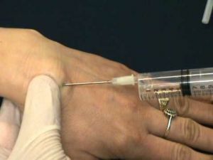 length. That is, all excess accumulated fluid is removed without cutting the skin. Thus, this method can also be classified as conservative. It is effective only in the early stages of the process.
 - With the advanced form of education, it is advisable only to use surgical intervention .Skin covers that are subject to tissue are dissected - access for surgical intervention is formed. Complete removal of the tumor along with its capsule and the attachment of healthy tissues to the subcutaneous fat are performed. Then, a seam is applied to the skin. As a rule, there is a scar.
Laser removal - an effective innovation
Now there is a modern method of removing the ginglion using laser technology.
The laser cuts the skin and the subsequent layers, removes the formation and sutures. After the operation, there are 2-3 points, which eventually disappear almost completely, which is more aesthetic.
The popularity of this method is also due to the fact that healthy tissues are practically not affected. And the occurrence of relapses is reduced to zero.
All types of surgical intervention are conducted under anesthesia, usually general. Therefore, do not be afraid that you will be hurt. The rehabilitation period is also not very large, only about 10 days.
Possible complications of
Such great importance to these seemingly trivial processes is due to the high number of complications.
Like any process of neoplasm, even benign, it can undergo malignancy - a transition from benign to malignant.
Preventive measures
 Prevention of ganglion development is the dosing of physical exertion. It is necessary to protect the joints from mechanical permanent trauma.
At all times the age of a person is determined by the state of his hands. If the hands were well-groomed, neat and beautiful, it was believed that this man is beautiful.
So follow the beauty and health of your hands, do not let any disease disturb your habitual way of life and affect happiness. Do not pull with an appointment with a doctor, it's better to make sure once again that everything is fine, than regretting what was not done. Be always healthy and beautiful!