The osteochondropathy of the tuberosus and apophysis of the calcaneus was first described in 1907 by Haglund. This disease is mainly found in girls 12-15 years old. The pathological process affects one or both extremities.
The disease is characterized by the appearance of aseptic necrosis of spongy bone sites, which are under the influence of increased mechanical stress.
Most often the osteochondropathy of the calcaneus occurs in children. Usually the disease is benign and does not affect the function of the joints and the general condition of a person.
Quite often the disease passes without treatment, as evidence of the transferred disease there are only deforming arthrosis.
Contents of the article
- Pathogenesis of the disease
- Causes of the pathology
- Diagnostic techniques
- What treatments exist?
- Surgery
- Conclusions
- Video: Surgical correction of foot deformities
Pathogenesis of the disease
The pathogenesis of the disease is not fully understood. It is believed that the osteochondropathy of the calcaneus is a consequence of local circulatory disorders that lead to a decrease in the nutrition of the adjacent tissues, which is the starting point of the development of the disease.
There are five stages of the development of the disease:
- aseptic necrosis( necrosis);
- fracture and partial fragmentation;
- resorption of dead bone tissue;
- repair( recovery);
- inflammation or deforming osteoarthritis, in the absence of treatment.
 Find out why the leg in the hip hurts from our material - the causes and illnesses that cause this kind of pain.
What you need to know before applying Voltaren gel - instructions for use, the pros and cons of the drug, indications for use and other useful information about the drug.
Causes of pathology
Researchers suggest that pathogenic factors such as microtrauma, increased stress( running, jumping), tension of the tendons of muscles attached to the calcaneus, endocrine, vascular and neutrophic factors cause osteochondropathy of the calcaneus.
Diagnostic techniques
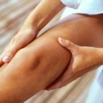
A characteristic feature of the disease is that pain in the area of the buttock bone appears only when it is loaded or when it is pressed, 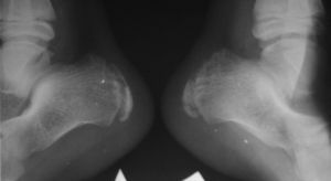 pain is absent.
This feature allows distinguishing this disease from bursitis, periostitis, osteomyelitis, tuberculosis of bones, malignant tumors.
Above the hillock of the calcaneus there is a swelling without reddening and any other signs of inflammation.
Most patients note the appearance of skin atrophy, moderate soft tissue edema, increased skin sensitivity on the plantar surface of the foot in the calcaneus. Quite often atrophy of the calf muscles.
X-ray examination determines the presence of the defeat of the apophysis of the calcaneus, which manifests itself by loosening the structure of the bones and cortex under the apophysis.
For pathology, a characteristic feature is the presence of shadows of dead bone areas displaced to the side, as well as the unevenness of the contour of the bone surface will be more expressed than normal on a healthy leg.
What treatments exist?
 Conservative treatment for this pathology is not always effective. But still, it's better to start with him.
In case of acute pain syndrome, a rest is prescribed, immobilization is carried out with a plaster bandage. For anesthesia, soft tissues in the heel area are chewed with novocaine.
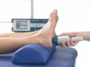
Carry out physiotherapy procedures: Novocain electrophoresis with analgin, microwave therapy, ozocerite applications, compresses, baths.
Assign medications such as pyrogenal, brufen, B vitamins.
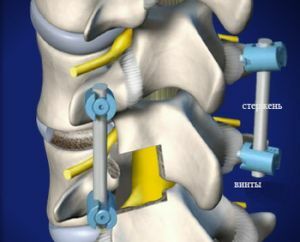
Surgery
If conservative therapy is ineffective, an operative intersection of the tibial and subcutaneous nerves with branches leading to the heel is resorted.
This allows the patient to get rid of unbearable pain, and also gives the opportunity to load heaps of heel bones when walking without fear.
Conclusions
In case of timely and correct treatment, the structure and shape of the calcaneus are fully restored.
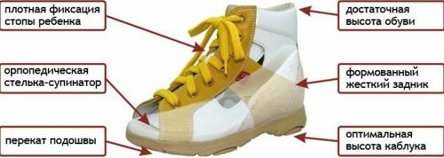
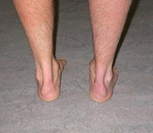
If you do not detect the disease in time or use irrational treatment, the increase and deformation of the calcaneal calcaneus will remain forever, which will make it difficult to wear ordinary shoes. This problem can be solved by wearing special orthopedic shoes.
Video: Surgical correction of foot deformities
Due to many leg diseases, the foot can be damaged. On the video shooting of surgical intervention in order to save the patient from pathology.