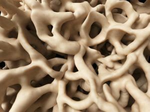
 The recovery time of a bone after a fracture can be from 4 to 6 months, depending on the complexity of the pathology. Throughout the time with the help of roentgenology there are signs of splicing.
In rare cases, this time is not enough for rehabilitation, and this is a sign of a delayed consolidation.
In case of fracture, fragments can be used not only for external but also for internal fixation, which in turn helps to adapt to bone fragments.
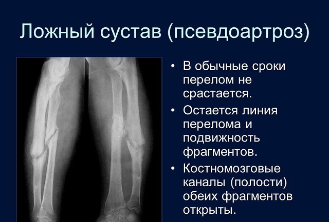
After healing, the fracture line is practically smoothed out and becomes invisible. But, in due course, it can expand a little, and in the intervals there is a bone irritating callus, devoid of precise contours, which is called a false joint or pseudoarthrosis.
Contents of the article
- Primary and secondary pseudoarthrosis
- What can be associated with the development of the disorder?
- Features of the clinical picture
- Objectives and diagnostic methods
- Treatment of violation
- A couple of words about the prevention of
Primary and secondary pseudarthrosis
To date, there are several types of reparative bone regeneration:
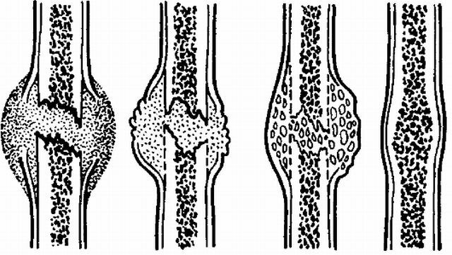
- The primary occurring in the shortest possible time. Splicing is effected through primary osteogenesis. In this case, between corpuscles, corn is formed - intermediate. In general, this kind of pathology occurs with compression or
 punctured fracture. If the fragments are fixed reliably and the reposition is ideal, then the diastase will not exceed 100 m.
 - If the bone grows together without a gap and only along the vascular canals, this kind of variety is called - a primary-delayed splice .In this case, only a partial splice occurs. For complete, interosseous intergrowth, resorption of the edges of the fragments is required.
- This kind of pathology implies the formation of several varieties of corns - endostal, periosteal and parasal. In this case, the fixation of debris is due to corns and is not performed by a specialist. With secondary splicing, the healing process can significantly increase.
What can be associated with the development of the violation?
As already mentioned, there is a congenital and acquired pseudoarthrosis. In this case, the congenital is extremely rare and is caused by intrauterine disruption of bone tissue formation or genetic disorder.
A fake joint of an acquired character can develop under the influence of many factors. The most common are the following provocateurs of the pathology:
- the formation of callus is due to a variety of disruptions;
- if there is diastase between the bone fragments and a displacement is observed;
- in some cases this may be the result of a violation of osteosynthesis;
- if the prescribed physical exercises are not properly performed or are inconsistent with the case;
- pseudoarthrosis develops upon attachment of a secondary infection;
- affect the formation of impaired blood supply.
In addition, incomplete fractures and pseudoarthrosis can be formed as a result of the following factors:
- as a provocateur of pathology may become a metabolic disorder( often it occurs in people prone to fullness);
- disorder may result from diseases related to endocrinology;
- infectious diseases;
- disorders related to blood supply, especially with large blood loss;
- in the case of multiple fractures;
- pseudoarthrosis develops in cases of innervation disorders;
- with combined lesions.
Features of the clinical picture
Symptoms of pseudoarthrosis:
- Motility is observed in places where it was not previously .This may change the direction of motion or increase the amplitude. Such manifestations in a healthy person are impossible. To date, there are cases when the arm or leg could rotate 360 degrees.
- A clear manifestation of the disorder is the limb shortening .The difference before and after, can be about 10 centimeters.
- Pathology also affects the muscles of the .There is a decrease in strength.
- About the disease may indicate loss of normal functioning of the limb .Especially it is manifested with the load, because in this case it is impossible to stand, walk and lean on the affected leg.
- Problems with the functioning of true joints .This is due to the lack of proper load.
Objectives and methods of diagnosis
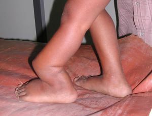
On the photo pseudarthrosis of the femur
In the diagnosis of pathology, it is important to take into account not only the collected clinical data, but also other factors. It is very important to take into account the estimated duration of bone splicing.
As soon as the deadline is passed, the status of the damage will be clear. If the result exceeds the established time by several times, the specialist diagnoses the formation of pseudoarthrosis.
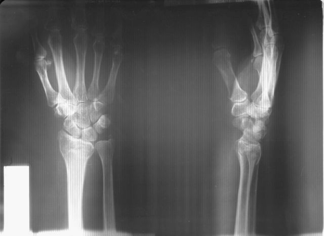
In addition, the patient is necessarily made X-ray in two projections. This action is necessary for the completeness of the picture, which will draw appropriate conclusions, and prescribe a treatment.
The fracture is established if the X-ray showed:
- complete absence of callus, which performs a connecting role;
- if the ends of the fragments are visible, and their smoothed, conical or rounded shape indicates a progression of the false atrophic joint;
- infestation of cavities of the ends of fragments.
If the patient develops pseudoarthrosis, the end of the fragment will have a hemispherical shape, which is more like a joint head. At the same time, the other end may be slightly concave. In this case, the x-ray clearly shows the formed articular gap.
If it is necessary to clarify the intensity of bone formation, an additional radionuclide study of the affected limb can be performed.
Treatment of a violation of
Pseudoarthrosis can have only one treatment option - surgical intervention. And the sooner the pathology is identified and eliminated, the painless the operation will be and the faster healing. 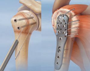
If hypertrophic pseudoarthrosis develops, then the specialist has the task of fixing the fragments. Various methods can be used for this, one of which is osteosynthesis.
At the moment when the fragments cease to move relative to each other, the process of recovery begins, which will give the first significant results after a few weeks.
If there is a development of atrophic pseudoarthrosis, there is a need to remove all sites that are poorly supplied with blood or do not receive proper treatment. In this case, the fragments must be connected, immobilized and left in this position until complete healing.
After the operation, the patient is assigned a course of massage and physiotherapy, which is aimed at restoring the proper functioning of the limb, blood supply and muscle strength.
Pathology, if left untreated, can lead to improper functioning of the limb or even complete loss of mobility.
A few words about the prevention of
Currently, there are no preventive measures for pseudoarthrosis. If we are talking about the acquired type of pathology, then the only thing that a patient can take care of is a cautious attitude to one's own body and timely treatment of a fracture.
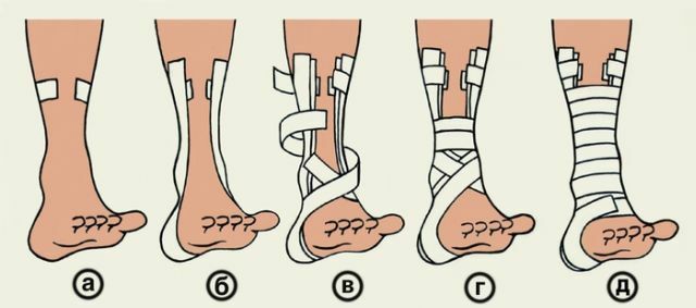
In addition, it is necessary to monitor the immobilization of the affected area. During the restoration of the limb, gypsum and other fixation methods are only removed by a specialist, it is not recommended to do it yourself unless such instructions have been received from a doctor before.
If before the time to remove the fixative structure, pain, may not follow, but there remains a great chance of incorrect or incomplete fusion of the bone, and with it the likelihood of developing pseudoarthrosis increases.