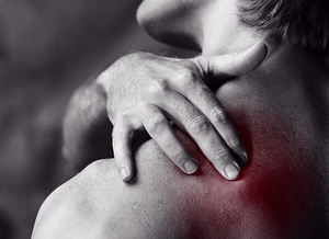
Back pain is one of the most common unpleasant symptoms in a person.
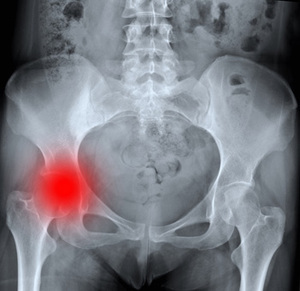
It ranks second in the frequency of seeking medical help after respiratory viral diseases. The cause of low back pain is spondyloarthrosis of the lumbosacral spine.
The lumbar region consists of five massive vertebrae. They are constantly experiencing high loads, because they provide a direct position of the trunk. Therefore, this department is prone to various degenerative diseases of more often than other .
Spondylarthrosis, is just one of such degenerative-dystrophic diseases.
In this pathology, both intervertebral joints between the vertebral bodies and the facet( small ridges) small joints are affected. At the same time, all the elements of the joint are destroyed: capsule, subchondral bone, cartilage, ligaments and muscles.
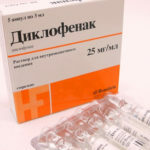 For the removal of pain, doctors recommend Diclofenac in pricks. Read the contraindications.
On what other injections are prescribed for osteochondrosis read the link.
Contents of the article
- The causes of spondylarthrosis
- Developmental anomalies
- Trauma
- Instability of the vertebra
- As spondylarthrosis develops
- Clinical picture of the disease
- Diagnosis of spondylarthrosis
- Treatment of patients suffering from spondylarthrosis
Causes of spondylarthrosis
In 80% of cases, the disease develops in the elderly. In young people, such changes usually develop after 25 years. There are three main reasons for the development of the disease.
Developmental anomalies
Congenital spine anomalies. This, as a rule, the presence of the sixth lumbar vertebra( lumbalization of the first sacral vertebra) or, conversely, sacralization - the fusion of the fifth lumbar vertebra with the sacrum.
The "transitional vertebra" is the most common cause of back pain, especially if it is an incomplete or one-sided sacralisation, that is, when on one side the transverse process is significantly enlarged and connected to the sacrum. The axis of movement here is displaced, and the tension of muscles and ligaments on the opposite side can be caused.
Similar anomalies can be a contraindication to certain professions. Pathology is clearly visible on X-rays.
The asymmetric position of the arcuate joints also plays an important role. Lumbar facet joints are located sagittally in the first two vertebrae, in others - almost coronary - parallel to the coronary suture.
Sometimes the sagittal position of the facet joint is on one side, and the coronary position on the other. This is predisposing factor for additional load on these joints during rotation.
Trauma
Injury develops aseptic inflammation in the joints of the spine. Edema of the tissues causes compression of the nerve roots, with pain. Violation of innervation leads to a violation of blood circulation in this area and to dystrophic changes in tissues.
The cartilage is heavily loaded with a traumatic subluxation of the facet joint.
Instability of the vertebra
This is an independently occurring pathology in which the lumbar vertebra is displaced forward or backward with respect to adjacent vertebrae. Most often, the forward displacement occurs when the slopes are forward.
Also from the causes of the development of spondylarthrosis can be noted scoliotic deformity, resulting in an uneven distribution of the load on the joints of opposite sides of one vertebra, which leads to asymmetric displacements and increased wear of the joints of the spine.
How spondyloarthrosis develops
The pathogenesis of the disease is not fully understood. Spondyloarthrosis is associated with the development of degenerative processes in the facet joints of the spine.
The trigger mechanism is usually considered to be frequent traumatization of these joints. In addition, the appearance of degenerative change in excess weight and the inheritance of .Under the influence of loads, the elastic pulp nucleus gradually loses its properties and ceases to fulfill its damping function.
In the conditions of pathological mobility( instability) of the vertebral segment adjacent bodies of vertebrae and joints are affected, so spondyloarthrosis develops. The joints are joined together by muscles and ligaments, under the influence of pathological impulses, they cease to fulfill their inherent functions, and the scoliotic deformation , characteristic of spondylarthrosis develops.
Clinical picture of the disease
The main symptom of spondylarthrosis is the pain that appears when moving and changing the posture. This pain passes in rest. As the disease progresses, there may be a morning stiffness in the lower back, which usually lasts no more than an hour.
Pain in spondyloarthrosis is local in nature and does not give up in the leg or groin, as with the intervertebral hernia. But with the progression of the process, compression of the nerve roots may occur, then the clinical picture of radiculitis develops with numbness and pain in the lower limb.
Spondyloarthrosis is most commonly developed in the lumbar spine. This is due to the presence of anatomical prerequisites in some patients - hyperlordosis and anterior pelvic incline.
Diagnosis of spondylarthrosis
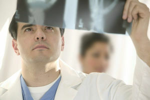
• Clinical data.
• Radiographic study.
• CT or NMRT.
Treatment of patients suffering from spondylarthrosis
Treatment of this pathology is due to the stage of the disease. 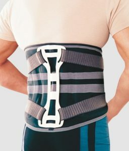
Outside the exacerbation of , a massage is prescribed, therapeutic gymnastics aimed at strengthening the muscular corset and visiting the pool. The development of the muscular skeleton of the spine allows to preserve the functional mobility of the spine for many years.
During the exacerbation of , anti-inflammatory drugs( NSAIDs), central muscle relaxants( midocals), acupuncture and traction( spinal traction) are prescribed.
When the reflex muscular-tonic pain syndrome develops, it is recommended that perform isometric exercises of with a careful transition to counter-acting exercises.
In older patients, , and if there are contraindications to performing muscle strengthening exercises, physiotherapeutic treatment is prescribed. To reduce pain, in addition to medicines, sinusoidally modulated currents are used, magnetotherapy, ion-galvanization with anesthetics, phonophoresis with glucocorticoids to stop inflammation and edema, massage and therapeutic gymnastics.
To restore the structure of the cartilaginous tissue, the preparations of the chondroprotective series( Dona, Chondroitin, Structum) are now widely used. These drugs not only stimulate regeneration in the cartilaginous tissue, but also slow the degenerative processes, contributing to the restoration of joint function.
The drugs have anti-inflammatory and anti-edematous effect, as well as restore the phosphorus-calcium metabolism. Their use of is justified in the early stages of , as it prevents the development of many complications.
A more pronounced effect is achieved when is treated with ultrasound with chondroitin .The drug under the influence of ultrasound penetrates more intensively into the affected tissues, in connection with this there is a more pronounced therapeutic effect.
The medical plaster Nanoplast forte has shown good results in the treatment of spondylarthrosis.
is installed and applied in our time.
It is most often recommended to install an InSpace spacer manufactured in Switzerland.
It is established by puncture through a puncture of soft tissues in the lumbar region under X-ray control. In this case, a special tool is carried out in the interval between the spinous processes and expands it, after which an implant of the appropriate size is installed here. After the implant is compressed, the achieved width of the interstitial space is retained. This allows to return to all nearby structures a normal anatomical relationship.