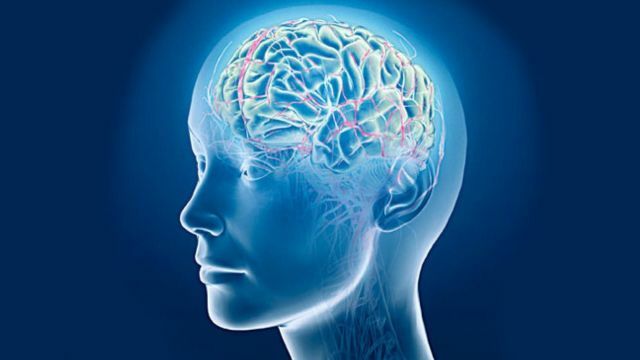
 In order for the brain to function in normal mode - without downtime and overloads - the blood circulation in it should be adjusted to the accuracy of the clockwork mechanism. Therefore, in addition to the need for an influx of oxygen and glucose with arterial blood supplying it with food, the archival is also the outflow of venous blood, which carries everything that the brain not only no longer needs, but is simply dangerous - the poisons formed during "thought production".
Here and there is always an extraordinary wit of nature, surpassing the degree of simplicity and elegance of solving the problem, all conceivable engineering ideas.
Contents
- Features of the structure of the venous outflow system
- Dysrecirculation: when venous outflow is obstructed or disturbed
- Results in between and final
- There are many in the brains of the regions - the reasons for them are no less amazing!
- Particularities and Nuances: Symptoms and Signs
- Confirmation of Diagnosis
- How is VDC treated: Therapy Methods
- Problem Prevention
- And so that without consequences!
Features of the structure of the venous outflow system
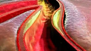
The system of venous outflow of the brain differs from that in other organs in that the veins do not accompany the arteries here. It is formed in the form of a ring structure, which has numerous anastomoses with an extracranial venous network, and uses for its needs existing in the brain formations, which gives considerable benefits.
First, the "tap water" of the brain is unremovable. It is formed not by soft ducts, but by sinuses - channels that pass epidurally - between two leaves of sickle shaped projections formed by the dura mater cerebri and creating the inner skeleton of the skull.
Sickles - like partitions inside a walnut - divide the inner space of the skull into several large cells that are not completely isolated from each other( and in the common "bedroom" each share of the brain has its own personal "cradle").
Simultaneously, they serve as ribs of rigidity - "rafters", which ensure the protection of the cranial roof from the threat of extrusion from the outside.
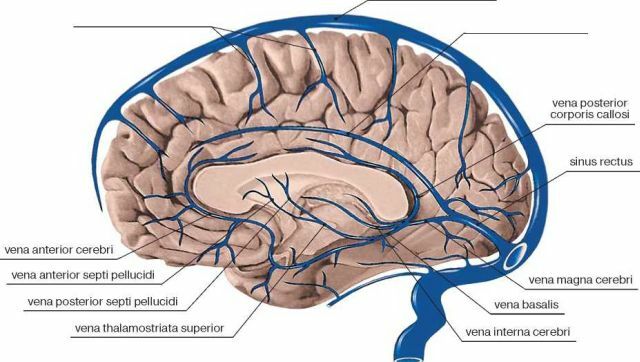
Veins of the brain
Secondly, the existing system of sinuses, using free edges of sickles - partitions between the parts of the brain - does not require the laying of any additional communications. Such an architecture resembling an aqueduct provides this structure with an enviable compactness.
Even more similar to the aqueduct has a large( sagittal) cerebral crescent. It forms a sine not only on the lower, free edge( the lower arrow sine), but also on the upper, attached to the bones of the roof of the skull from the inside( the upper arrow sine).
Lower sagittal sagittal sinus, "climbing" on the "horse" of the cerebellum whine, forms a short straight sine. The message of the latter with the superior sagittal sinus and two obliquely horizontal parietal-occipital( transverse), assuming paired temporal, forms a "cross", called the sinus drain, or the pulp of Gerophilus;its component part is also the occipital sinus.
In addition, the system includes:
- sigmoid sinuses - paired( available on both sides), serving as a continuation of the transverse, into which the lower stony sines flow;
- the upper rocky sinuses , which flow into the transverse ones;
- cavernous sinus is an extensive "delta" around the Turkish saddle( from the fusion of paired sphenoid-parietal sinuses and formed with the participation of transversely interspecific sinus - anterior and posterior), which has anastomoses with venous plexuses of the outer base of the skull.
Sigmoid sinuses, in turn, become the beginning of internal lobes.
Venous sinuses are manifolds where the blood is collected and discharged from the veins of the usual structure, both superficial and deep.
Surface structures( cortex and white matter of the brain) are served by short cortical veins of the subdural and subarachnoid spaces:
- by the upper anastomotic vein of Trolar;
- dorsal superior cerebral vein;
- superficial middle cerebral vein;
- the lower anastomotic vein of Labbe.
The pathway of blood from the deep brain zones( in particular, from the thalamus and basal nuclei, the tissues that form the walls of the ventricles and the vascular plexuses) lies:
- in the internal cerebral veins - paired veins, each formed by the fusion of a septal vein that collects blood inthe area of the transparent septum, and the vein of the thalamostriary;
- into the veins of Rosenthal ( also paired).
These two pairs of vessels behind the platen of the calloused body discharge blood into the galenic( large cerebral) vein, from where it passes the sinus straight, 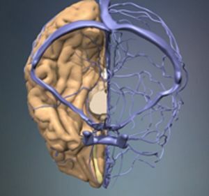 falls into the sinus pulp of Gerophilus.
The largest part of the venous blood from the surface of the brain is collected in the upper sagittal sinus, where it moves along it from front to back, while the blood from the deep sections of the brain takes a direct sine. The drain from the transverse sinus occurs in the sigmoid sinus located on the same side, below the yarrow hole becoming the inner yoke.
The venous blood from the basal parts of the brain is also produced in the cavernous sinus, where most of the blood is collected from the orbital regions and from the temporal lobes of the brain. Evacuation from the cavernous sinus is possible in two directions: partly through the lower and upper stony sinuses into sine sigmoid, in part by the withdrawal through the pterygoid plexus.
The blood does not necessarily leave the cranial cavity, leaving the inner vaginal veins. This can be done by means of a pterygoid venous plexus with discharge of blood into the viscarocarcinium( venous system of the facial part of the skull), and with the participation of emissaries - venous anastomoses in the thickness of the cranial roof bones that connect the sinuses of the dura with both diploic veins and veins of the outer regionshead.
Dyskirkulyatsiya: when venous outflow is difficult or disrupted
The venous network of the brain is a reflexogenic zone with a high level of nervous organization, responsible for the most important physiological processes, due to ensure the constancy of the blood supply to the brain.
"Dis-" - this means that the process is upset and out of control. When it comes to the disorder of circulation, this indicates a more or less significant imbalance of metabolism in the brain:
- oxygen;
- carbohydrate;
- water;
- fat.
As well as the increase in hypoxia and hypercapnia, venous and intracranial pressure, which leads to the development of cerebral edema.
Venous outflow disorder takes place in its path in 3 stages.
- In the latency stage , complaints are almost non-existent, clinical symptoms are not manifested.
- The period of cerebral venous dystonia is characterized by paraclinical changes, the symptoms are few and do not interfere with life.
- The detailed picture of venous encephalopathy requires specialist intervention, because it is already expressed by persistent organic microsymptomatics.
According to the authoritative opinion of M. Ya. Berdichevsky, the violation of venous outflow exists in two main forms: 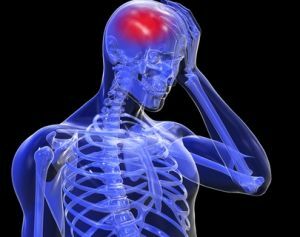
- With the primary form of is the growth of venous tone disorder, the basis for the development of venous circulation is chronic nicotine or alcoholic intoxication, hypertensive or hypotonic disease, venous hypertensioneither endocrine pathology, hyperinsolation or CCT.
- With , the stagnant form of , a violation of the flow of venous blood from the cranium is caused by mechanical causes, which leads first to a slowing of the venous circulation, then to stagnation of the venous blood, and eventually to the edema of the brain.
Intermediate and final results
Venous blood flow disorders may have a variant:
- of venous stasis;
- of encephalopathy of venous origin;
- hemorrhages of venous etiology;
- thrombosis of veins and sinuses;
- thrombophlebitis.
Some authors adhere to the classification of EZ Neimark distinguishing both the cranial vein structures failure, and the disorders of the veins of the main type and the disorder of the co-genesis, dividing each type of disorder into:
- acute and subacute , including variants of venous hematoma formation andhemorrhages( intracerebral, as well as subshell) on the basis of thrombosis or intracranial veins, or sinuses, as well as due to phlebotrombosis of veins and sinuses, or their phlebitis or thrombophlebitis;
- chronic cases caused not only by encephalopathy of hypertonic and atherosclerotic, but also by encephalopathy of the venous.
Chronic venous insufficiency( in the form of encephalopathy) can occur in the form of symptom complexes, leading to the development of a number of pathological states of the brain and nervous system:
- asteno-vegetative;
- pseudo-tumorous hypertensive;
- psychopathological;
- stroke;
- polymorphic.
And can cause:
- bettoleptic;
- syndrome of terminal and preterminal convulsions.
A lot in the brains of the regions - the reasons for them to hit no less!
Localization of the affected area of the brain, its nature and depth depend on the causes of the development of venous dyscirculation, and the symptoms that express it also "dance" from them.
To frequent reasons for a disorder of venous outflow from the brain, one should consider:
- pulmonary, or cardiac, or pulmonary-cardiac failure;
- is the transmission of strategically important extracranial veins, such as the inner jaw, the nameless, the upper hollow;
- malignant or benign tumors of the skull and brain;
- ЧТМ;
- thrombosis of veins or sinuses of the brain;
- craniostenosis and edema of the brain, leading to compression of the veins;
- asphyxiation of newborns;
- as well as the cause of suicide or forcibly applied - hanging.
Most often this is due to thrombosis of veins of different depths or venous sinuses of the brain( and the clinical manifestations of phlebothrombosis will not differ in any way from those in thrombophlebitis).
Particularities and nuances: symptoms and signs
 The clinic of thrombosis of superficial cerebral veins usually combines neurological symptoms with characteristic signs of inflammatory, especially infectious, lesions( with hyperthermia, an "inflammatory" response from the blood and liquor).
Often, the disease "debuts" a headache with nausea and vomiting, a violation of consciousness( almost always with psychosomatic arousal) serving as a backdrop for the development of focal brain symptoms( paralysis or paresis of limbs, aphasia, generalized or focal epitopes), the usual lability of which is explained by the movement of actionfrom the initially injured venous trunk to the adjacent one.
The ongoing studies result in demonstration of evidence of the above described symptoms: the detection of hemorrhagic strokes in one or both types of brain substance, subarachnoid or intracerebral hemorrhages, a picture of ischemia and cerebral edema;lumbar puncture ends with getting a hemorrhagic liquor.
In the vast majority of cases, thrombophlebitis of the veins of the brain surface accompanies the postpartum period.
Emphasis should be placed on the onset of cerebral symptoms in the presence of previously identified active foci of inflammation or thrombophlebitis of the extremities, on the appearance of cerebral symptoms both after the manufacture of abortion and in the postpartum period, as well as after the processes in the middle ear, in the nasal paranasal sinuses and after infectiousdiseases.
The general picture of venous sinus thrombosis, accompanied by a violation of venous outflow of the brain is quite typical: 
- a sharp headache;
- with characteristic "meningeal signs";
- marked edema of the skin of both the facial and the scalp;
- hyperthermia;
- different degrees of change in the state of consciousness( from the confocal to the coma).
In the study of the fundus there are clearly visible phenomena of stagnation and edema. In the analysis of blood - leukocytosis, in the cerebrospinal fluid( clear or xanthochromic) - a mildly expressed pleocytosis. Focal neurological symptoms suggest localization of the involved sinus.
The most common thrombosis of the sigmoid sinus complicating purulent mastoiditis or otitis is the characteristic soreness and swelling of the skin and soft tissues of the mastoid region, with increased sensations in both masticatory movements and head turns in the direction opposite to the one where the process developed,accompanied by significant septic phenomena.
If the process is thrown to a vein, the symptoms of nerve damage IX, X and XI appear on the side of the focus.
What is the manifestation of a cavernous sinus thrombosis, which is a frequent consequence of purulent inflammation on the face, in the orbit, in the ears, in the sinuses?
The appearance of indisputable signs of the difficulty of venous outflow in combination with the clearly manifested symptoms of the inflammatory process in the form of:
- periorbital edema or edema of the eyelids;
- chemosis;
- of increasing exophthalmos;
- stagnant picture of the fundus with signs of optic atrophy.
The appearance of: 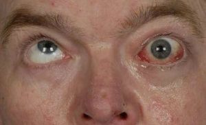
- of external ophthalmoplegia( due to the involvement of III, IV, VI craniocerebral nerves) is also possible;
- ptosis;
- disorders of pupillary reactions;
- tarnishing of the cornea;
- pain in the forehead and eyeball area( due to involvement of the upper branch of the trigeminal nerve);
- sensitivity disorders in the area of the supraorbital nerve.
Severe cavernous sinus thrombosis can occur with a two-sided variation, when the process can spread to adjacent sinuses.
It is possible and aseptic flow of thrombosis of the cavernous sinus, which developed as a result of hypertension and as a result of atherosclerosis.
The thrombosis of the upper sagittal sinus differs in the variability of the clinic, which depends on the cause, the rate of increase of thrombosis, the place occupied by it on the scale of the sinus, and the extent of involvement in the pathology of the veins that make up its basin - this is an extremely complex septic case of thrombosis.
Thrombosis of the upper sagittal( longitudinal) sinus is characterized by blood overflow and vein tortuosity:
- century;
- of the base of the nose;
- temples, forehead and crown with a massive edema of the whole region( picture of the "head of the jellyfish"),
A, in addition, frequent nasal bleeding, painful when trying to perepututirovat parasagittal area.
Neurological symptoms are based on signs of intracranial hypertension, as well as frequent( beginning on the feet) convulsive seizures;Perhaps the emergence of lower paraplegia with enuresis or tetraplegia.
Other types of thrombosis of the sinuses include the mystical( on the basis of exhausting diseases in old people and infants) and infectious thromboses of both cerebral veins and sinuses, which can be complicated by the development of encephalitis, purulent meningitis, and brain abscess.
Diagnosis confirmation
The diagnosis is confirmed by a method capable of confirming the truth of the alleged pathology and giving an exhaustive picture of 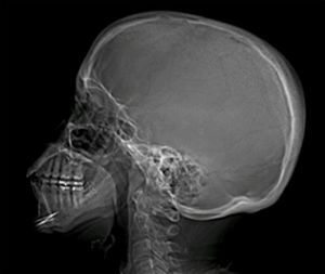 of the state of the veins of the brain( especially the hilar veins).
The most commonly prescribed MRI.
Other valuable research methods are:
- radiograph of the skull;
- phlebography;
- examination of the fundus.
How VDC is treated: therapy methods
At a time when the disease has only just begun to manifest itself, it is enough to adjust the mode of work and rest.
In case of persistent continuation of the venous outflow disturbance, it is worthwhile to seek the help of a specialist-a neuropathologist-doctor who will recommend an adequate medication for the condition.
For the most effective care, both the general condition of the patient and its particularities are evaluated( for example, with the concomitant varicose process, the use of disaggregant drugs, for example, aspirin) will be expedient.
Most often, if there is a violation of the venous outflow of the brain, the use of venotonic agents is recommended:
- normalizing blood circulation;
- improving vascular function;
- giving veins elasticity;
- strengthening walls of blood vessels;
- contributing to their adequate permeability;
- removing edematous phenomena;
- preventing the development of inflammation and fighting with existing ones;
- boosting the tone of the body.
 All this can significantly improve the "living standards" of the veins of the brain.
This group includes: Anvenol, Venoplant, Escuzane, Venen-gel and others.
In order to increase the vascular wall resistance, periodic injecting courses of nicotinic acid and pyridoxine are used.
For the elimination of brain symptoms serve as long-term drugs-nootropics: Fentropil, Glycine.
From non-drug therapies, massages and self-massage courses( conducted after training with a specialist), especially the neck area, are strongly recommended.
Prevention of
problems No less than in treatment with developed pathology, the body needs and prevention of venous outflow problems - regular self-diagnosis.
Urgent examination with a neurologist and ophthalmologist is necessary with the necessary research when:
- is a dull headache, worse with head movements;
- edema of the lower eyelid;
- cyanosis of the cheeks, lips, nose;
- a hum in the head with a maximum of manifestations in the morning;
- of pronounced meteorological dependence;
- fainting, either dizziness or blurred vision, not to mention mental disorders and epileptic seizures.
The prevention of venous outflow disorders from the brain is also the maintenance of optimal sleep,  wakefulness, care for proper nutrition, eradication of the usual intoxications and other harmful traditions.
Other valuable methods of influence on the body to improve its condition are:
- various relaxation techniques;
- application of herbal medicine;
- taking a contrast shower;
- using yoga.
And so that without consequences!
Not looking after your health, or continuing to stubbornly cling to your old habits and lifestyle( with the diagnosis already established), you risk losing not only your health, but life as well.
Because cerebral hemorrhage, the cause of which can be venous disgemia( the same as discirculation), can lead to both a wheelchair and a place in the cemetery.
Relatively "sparing" consequences are aphasia, mental disorders, the appearance of convulsive seizures and the development of paralysis or paresis in the limbs.