
Breast cancer in women are a vulnerable point for the development of education (benign or malignant), especially during menopause, when there is a decrease in the production of female hormones. Tumor, detected at an early stage is more amenable to therapy with a possible full recovery.
But since the early stages of disease without the characteristic symptoms develop, for the early detection of cancer or cysts need to undergo an ultrasound or mammography breast annually.
The content of the article:
-
1 Mammography: principle, types of equipment
- 1.1 Analog and digital mammography
- 1.2 tomosynthesis
- 1.3 Ductography (galaktografiya)
-
2 Under what conditions need a mammogram?
- 2.1 Preparation for studies of breast
- 2.2 pros techniques
- 2.3 Minuses
-
3 Ultrasound examination: the principle of equipment
- 3.1 Simplicity and ease of
- 3.2 When ultrasound is necessary
- 3.3 pros techniques
- 3.4 Minuses
- 4 Is it possible to do both at once studies of breast?
- 5 A comparison of the accuracy, informativeness, safety, comfort, number of disadvantages, price
- 6 Which method is better?
- 7 Video on the effectiveness of breast ultrasound and mammography
Mammography: principle, types of equipment
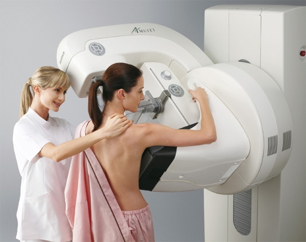
Mammography - a non-surgical method of breast examination with the use of X-rays. To pass the examination of the breast is placed between the work plates the X-ray apparatus (woman in the period procedure is standing or sitting) and is fixed.
This allows to align the gland tissue thickness and to avoid distortion of the results. Abdomen covered lead apron, to prevent damage to organs beams arranged in the abdominal cavity. The state of the mammary glands is estimated in the projections, which is obtained by passing radiation through the tissue in direct and oblique directions.
The principle of operation is based on the fact that different density tissue differently passes X-rays. As a result, the presence of seals or salt deposits in the breast, the bright spots are displayed in the picture. A tumor or voids are visible in the dark formations tissues.
Analog and digital mammography
When received analog mammography images recorded on the film in the form of individual images (as chest x-rays), and when digital data is displayed on special media.
The latter procedure is released high-resolution images and the ability to zoom, allowing you to more accurately determine the state of the glands. There is the possibility of obtaining copies of the information to digital media. It used when necessary to clarify or diagnosis in different clinics at the same time.
tomosynthesis
A leader in mammography as it allows to obtain three-dimensional images of the mammary glands. During the procedure, with the X-ray tube are rotated around the breast.
As a result, sections (images) obtained at different angles. The information obtained is processed by computer and displays a 3D image on the screen. The downside of the procedure is the increased radiation dose.
Ductography (galaktografiya)
The method used for checking the condition and patency of mammary ducts. In conducting the contrast agent used. In his passage through the ducts is determined by their physical condition (thickness, presence of tumor formation and the existing abnormal narrowing). Upon detection of abnormalities require further examination.
Under what conditions need a mammogram?
Breast ultrasound or mammography (better to consult a gynecologist or breast physician before the procedure) - methodology breast exam, the selection of which is determined depending on the purpose (preventive examination or the presence of symptoms pathology).

Mammograms produce subject to the following deviations:
- appearance of pain in the breast without a provoking factor (onset of menstruation, pregnancy, trauma);
- presence of seals on palpation (may be carried out at home by the mammary glands, gynecological or mammologa);
- increasing the size of one of the glands without a cause;
- occurrence of secretions of the mammary ducts (not during breastfeeding grudnichka);
- varying a nipple or peripapillary region (color, shape, or drawn into a different strain);
- redness or dryness of the skin or epidermis coarsening without provoking factor;
- a sense of "preserver" breasts are not related to pregnancy, PMS or feeding;
- to examine changes in the breast in the presence of malignant and benign tumors;
- to identify the location of the tumor prior to surgery;
- the presence or onset of menopause endocrine abnormalities;
- predisposition to cancers;
- breast examination after tumor removal by surgery.
Once every 24 months to pass a mammogram is recommended as a preventive measure.
It is important to carry out mammograms prohibited when:
- pregnancy;
- breastfeeding;
- open chest injuries.
If the procedure is necessary under these conditions, it is only with the permission of the treating specialist. 40 years mammography is ineffective due to the increased density of breast tissue.
Preparation for studies of breast
For effective results mammography is required:
- undergo the procedure 5 through day 14 of the menstrual cycle. In this phase of edema and swelling of the glands is at a minimum. In the absence of menstruation these restrictions are absent;
- on the breasts and armpits be absent cosmetics (powder, essential oil perfumes, deodorant composition). They distort the results of the survey;
- if any implants or scars from breast surgeries. This should be forewarned specialist;
- eliminate alcohol, coffee and chocolate for 3 days prior to the procedure. They influence on hormonal balance, and also have a widening effect on blood vessels, a hundred and distort the results of the survey.
During the procedure you want to relax the muscle tissue in the mammary glands. If the survey is planned to determine the dynamics of the chest, then you need the results of previous reports.
pros techniques
The advantages of the survey method of mammography include:
- It can detect even small deviations in the tissues of the mammary glands and ducts;
- from the images we can determine the place of localization of formations and their characteristics (shape, density, size);
- Detection salt concentrations in the breast is only possible with mammography;
- It is the most effective method for the detection and examination of cystic formations;
- one can determine the slightest change from the images (when examining the dynamics of pathology).

Results are arms patient. you can apply to various medical facilities for diagnosis.
Minuses
Breast ultrasound or mammography (better determine the type of inspection is not only advantages, but also disadvantages) - techniques which have disadvantages as during the procedure, and in the performance data.
In mammography, these include:
- the procedure is the primary, further examination is required for accurate diagnosis;
- to 40 years mammography give skewed results (due to high density breast tissues);
- calculate the required days of the menstrual cycle (as for a free inspection record on such days may be omitted);
- slight discomfort due to compression gland plates;
- the body receives radiation which has a negative impact on its activities. Therefore, the procedure is not recommended to carry out more frequently than once every 12 months;
- it is impossible to detect changes in the lymph nodes.
In the presence of the implant performance of mammography is low. Also, the breasts are too large or very small, can cause inefficiencies procedure. In rare cases, may produce false results. It depends on the technological characteristics of the equipment used and klassnosti specialist who conducts mammograms.
Ultrasound examination: the principle of equipment
Ultrasound is also non-invasive method of determining the conditions of mammary glands. To conduct ultrasonic waves are used procedure. The principle of the method is based on the ability of different density tissue differently, display, diffract or absorb the rays. But ultrasound is effective only in benign tumors, or entities with liquid contents.
In the presence of cancer cells efficiency is low.
The procedure is performed in the prone position. Specialist handles mammary gland specific composition and examine it, using sensor. US data are displayed on the monitor. With the use of the Doppler effect can identify the status and permeability of the blood vessels in the gland.
Simplicity and ease of
Breast ultrasound highlighted the simplicity and convenience in the survey. Therefore, for the diagnosis of pathologies are not malignant, the method survey is considered better when compared with mammography.

To carry out ultrasound is required:
- be screened from 5 to 10 hours from the onset of menses. Then productivity will be higher procedures. But an urgent survey data limitations are ignored, if necessary;
- the skin should be clean. The use of deodorants, aromatherapy sprays and other cosmetics on the day of the procedure is prohibited.
With the passage of the planning ultrasound to track the dynamics of therapy or educational growth, are required to provide data from previous procedures.
When ultrasound is necessary
Procedure for ultrasound breast examination shows the following pathologies:
- predisposition to the development of pathologies in the breast and in the menopause;
- determining the status of the breast tissue in the presence of silicone implants. A state also implants themselves;
- pain in the glands of unknown nature;
- tracking the dynamics of treatment of cystic formations;
- the presence of uncharacteristic discharge from the breasts;
- detection of deviations in the glands during the childbearing or under natural feeding (mastitis, vascular status and the presence of abnormalities in the blood flow);
- determining the status of lymph nodes and breast tissue during the development of inflammation or trauma;
- Track the status of the breast after surgery;
- lump or swelling of tissues in the breast;
- change tone and appearance of the skin or nipple (darkening, peeling, deformation);
- chronic gynecological diseases or hormonal imbalance.
The procedure is recommended to take a preventive measure every 12 months. Age and other restrictions to conduct ultrasound available.
pros techniques
Breast ultrasound or mammography (the best tool for diagnosis is also determined by the quality of equipment and qualification of specialist) appointed individually by a breast.

Determining the status of breast tissue by ultrasound has the following advantages:
- studying tissue process is painless and harmless;
- suitable for any age and during pregnancy and lactation;
- You can detect changes in the lymph nodes and blood vessels;
- to determine the state of the prostate tissue in the presence of silicone implants;
- procedure, if necessary, may be repeated without sustaining time interval;
- It can be considered a pathology from any angle;
- prostate size does not affect the quality of the procedure;
- reveals metastases and the affected area;
- the procedure can be carried out in the presence of inflammation and internal injuries in the gland.
Ultrasound is used during the puncture of breast tissue. Mammography for this purpose is less informative.
Minuses
Before selecting ultrasound is recommended to get acquainted with the procedure cons:
- Assessment of prostate tissue depends on the experience of modern equipment and skilled conductive procedure, and they also issued conclusion;
- diagnosis made by ultrasound requires additional procedures to confirm or refute the type of pathology;
- to determine tumor cell procedure is ineffective.
The equipment does not allow to identify the presence of salt deposits in the chest.
Is it possible to do both at once studies of breast?
Breast ultrasound or mammography (better carry out procedures together) effective to detect pathologies depends on the type of education and qualification of the doctor conducting interpretation of the results. When deviations are detected by mammography may require confirmation of tumor localization by ultrasound, as well as carrying out a puncture with this unit.

If detected presumably Cancer tumor using ultrasound to confirm the additional assigned mammograms. Jointly conducted procedures increase the accuracy of diagnosis. It is important to bear in mind that mammography has age restrictions.
A comparison of the accuracy, informativeness, safety, comfort, number of disadvantages, price
Comparative analysis of mammography and ultrasound on the basic parameters:
| comparison items | Mammography | |||
| analog | digital | tomosynthesis | ductography | |
| Comfort | During the procedure the patient is in a standing or sitting position. Mammary glands are located between the X-ray plates, causing discomfort and sometimes pain. For the accuracy of the result is required to hold their breath. | Performed with analgesics, since the process of administration of contrast agent is painful (composition is introduced through the lactiferous ducts). | ||
| Security | Methods are safe, since there is no damage to the tissue. But with injuries this survey method is not applicable. During pregnancy, the procedure is not used. Breastfeeding needs clarification and the treating specialist. | When negligence specialist possible infection in the milk ducts. Pregnancy and breast-natural does not apply. | ||
| Accuracy depends on the percentage range of modern equipment | 90% | 90-95% | 95-99% | 90-95% |
| Informa-ciency | It lets define benign and cancerous, as well as salt deposits. When Ductography can track the operation of the milk ducts. | |||
| Advantages and disadvantages | Pictures can not be duplicated, to increase image quality. The minimum irradiation beams. | The information on the recording medium can be expanded, to increase or decrease the image and change the contrast for a sharper image. | The radiation is higher than in other methods of mammography. It allows you to get a 3D image with high precision localization of education. | Soreness procedure. The only method to determine the state of the milk ducts. |
| The cost (minimum of Russia in rubles). | 1500 | 2200 | 3200 | 1500 |
| Efficiency of up to 40 years | Ineffective, since the mammary gland consist of denser tissue, distorting the result of research. | |||
| Efficiency after 40 years | Efficiency of the procedure is high (when compared with the US). | |||
| Efficiency after 50 years | Efficiency is also high, but the risk of tissue damage is reduced by irradiation glands. | |||
| Efficiency after 60 years | More convenient to use, since there is no need of waiting the required period in the menstrual cycle of surveys. Also, damage to the tissues rays is minimal at this age. |
| comparison items | ultrasound |
| Comfort | The procedure is performed in a lying position and does not cause physical discomfort. |
| Security | The integrity of the skin is not broken. The probability of infection in the gland is missing. Allowed examination at gestation and lactation. |
| Accuracy depends on the percentage range of modern equipment | 85-90%. |
| Informa-ciency | In cancer treatment is ineffective, but to determine the state of the iron in the presence of silicone implants. It is also possible to determine the state of blood vessels and blood supply quality in the glands. Ultrasound to determine abnormalities in the lymph nodes. |
| Advantages and disadvantages | Does not reveal cancer. But the procedure is effective for determining benign at any age. The multiplicity of procedures is not specified. Allowed retested undelayed slot. Allowed to survey the presence of inflammation in the chest. |
| The cost (minimum of Russia in rubles). | 300 (using the Doppler 3500) |
| Efficiency of up to 40 years | It makes it easier to detect changes in the breast. |
| Efficiency after 40 years | It reveals tumors, but mostly benign |
| Efficiency after 50 years | Used to identify entities not oncological nature. |
| Efficiency after 60 years |
When passing ultrasound or mammography examination is required to give preference to the use of modern equipment and those of crusts, which confirm their qualification. On examination in the state institutions can not be selected, but then the procedure is free of charge.
Which method is better?
Choosing a breast examination is recommended to produce, together with the attending physician according to the available indications and subject to age limits. Passage both diagnostic procedures increases efficiency by up to 98%. It is important to determine the cancer is only suitable mammography, and in the presence of silicone implants need to select an ultrasound.

Mammary glands can be inspected in the home to detect visual abnormalities.
Further diagnosis is performed in hospitals on a fee or free of charge. Initially, most experts appointed by the passage of the ultrasound examination. If necessary, confirm or refute the need to diagnose cancer using mammography, which detects malignant tumors better.
Author: Svetlana Kotlyachkova
Video on the effectiveness of breast ultrasound and mammography
Learn more about breast ultrasound:
Fragment of the program "Live healthy" about mammography:
