Content
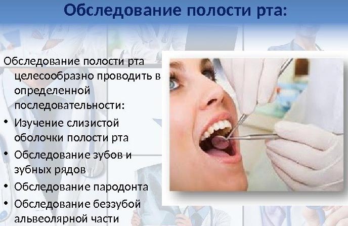
- Oral examination technique
- Primary symptomatic and visual examination
- Features of the
- Study of the architectonics of the vestibule of the oral cavity
- Periodontal assessment
- Assessment of the condition of the bite
- Assessment and registration of the condition of the teeth.
- Oral examination video
One of the most effective methods of dental examination is a complete examination, which allows the doctor to identify primary pathological processes, after which - to prescribe a high-quality treatment. The algorithm makes it possible to diagnose all components of the oral cavity, therefore this type of examination is especially important for dental health.
Oral examination technique
The beginning of the technique for conducting any examination, including the oral cavity, is characterized by a conversation in which the specialist determines the patient's main complaints, and also examines the anamnesis received.
In some cases, when collecting clinical information, the nuances and certain characteristics of the patient are clarified with his relatives and friends, as well as based on the patient's medical record.
The resulting data should be presented in relation to 3 main sections:
- any complaints from the patient, regardless of their objectivity;
- a full life history, including various methodological studies;
- a detailed history of the pathological processes present.
Any complaints from a patient that may affect health conditions are determined to be active even before the research process. This allows you to avoid in the future various complications in the form of incorrectly started treatment or surgery.
After that, you should find out the main factors affecting the occurrence of painful processes not only in the oral cavity, but throughout the body. Such a symptomatic picture makes it possible for the doctor to suspect the presence of dangerous inflammatory processes that indirectly affect the dental characteristics of the patient.
Incoming complaints can be characterized by both variety and localization in a certain area of the body. Often they are associated with painful sensations and other pathological processes affecting aesthetic and functional disorders of the facial or jaw region.
Primary symptomatic and visual examination
The history of the whole life is carried out based on the questioning of the patient about possible chronic or genetically determined diseases, including previous injuries or serious illnesses.
In the case of a child examination, questions are asked to the parents of the child, in which, first of all, it is recognized:
- the exact age of the child;
- hereditary predisposition that can affect degenerative changes in the bone or tissue structure of the oral cavity;
- the presence of chronic diseases in the mother of the child;
- gynecological and obstetric examination of the mother, including the number of births, pregnancy, and the presence of infectious or bacterial diseases during gestation. Also, the doctor asks the parent about possible toxicosis, dermatitis, bleeding and anemia;
- questions about the development of the baby, including weight and length at birth. Clarifies information about when the baby first began to hold his head, walk or sit. Also, the doctor learns about the type of feeding and taking medications in the first 1-2 years of life;
- external stimuli and living conditions, including the presence of a large amount of sweets in the diet;
- the level of hygiene and sanitary standards in the house;
- salient features of neuropsychic function, including head position at night, breathing rate and rate, and child's enthusiasm and academic performance.
In the presence of complaints that can cause various painful sensations, the nature of the disease is clarified. The child's history takes into account the words of the parents that indicate the presence or absence of dental care, the use of a brush and paste. Clarifies information about whether the child underwent dental treatment or any other pathologies in the oral cavity.
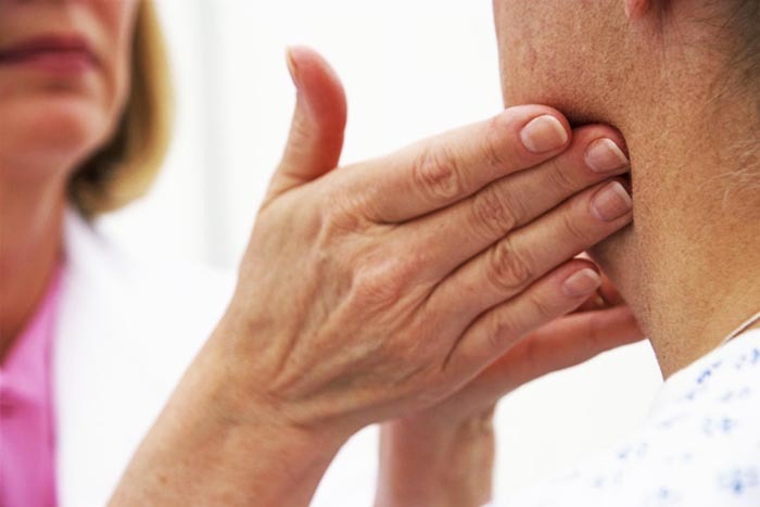
The doctor palpates the lymph nodes, which allows you to determine their condition. In the presence of probing, the size, mobility and consistency are revealed. Painful sensations are also tested.
The immediate dental status of the patient is determined based on the diagnosis of the configurational features of the face, including its skin, color, elasticity and the presence of a scar surface.
In addition, the presence of sinks or protrusions of the jaw, as well as other facial elements - the chin, lips, middle part of the face, is established. If possible, asymmetric features are identified that affect the growth of bone tissue.
Separately, the analysis of the shape, symmetry, size of the lower part of the jaw is carried out, the eyes, ears and nose are examined. Normally, the patient should not show any deviations when opening the mouth, without the presence of any displacement from the jaw muscles.
Features of the
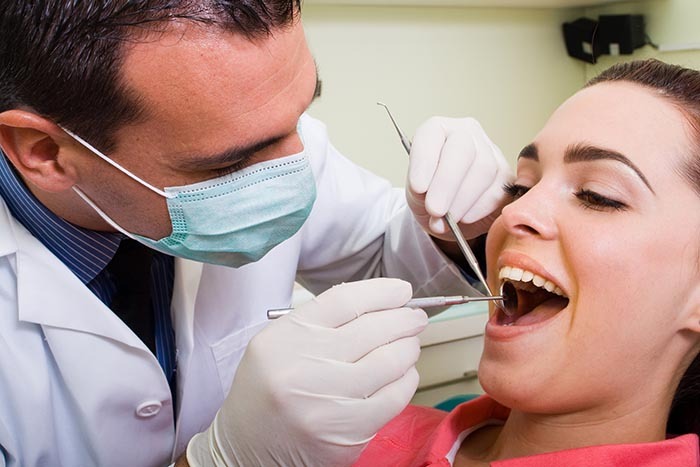
Examination of the oral cavity (the algorithm of which includes the study of all pathological conditions) is carried out in the dental chair. In case of diagnostics of children under 4 years old, they can be kept by the parent himself.
The patient can sit or lie on the chair. The dentist is most often located at the head of the person, which allows for good visibility and lighting. The diagnostic procedure is carried out using a dental probe, a mirror, and in some cases an air gun.
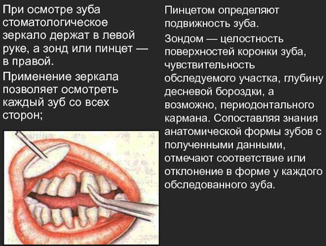
Since it is important in the study to achieve good focus on the object, the dental mirror can give an enlarged image of the surfaces of the teeth that are not accessible to normal vision. The location of the mirror is individual, depending on whether the dentist is left-handed or right-handed.
The dental mirror is held by 2 fingers, which are located at the base of the handle. In order to obtain a different projection of the oral cavity, the device tilts using the movement of the pendulum, and also rotates based on the element under study.

A dental probe is often used to remove unwanted objects that can interfere with the study, as well as to detailed assessment of mechanical damage or features of fillings, dental tissues, pathological complications and other properties of the oral cavity.
The probe is held with 3 fingers, while it is important that it should be perpendicular to the study area.
Despite the distinctive advantages of using sounding, such a procedure can be harmful in the event of:
- mechanical damage to the tissue structure, including unformed enamel, gums or fillings;
- infection with an improperly processed device, which is often accompanied by additional complications in the form of suppuration or complete loss of bone tissue;
- strong painful sensations due to the use of a probe in the study of open caries;
- psychological pressure of the patient, because the device itself looks like a needle.
Based on the above disadvantages, in modern dental practice, the probe is often inferior to another type of device - air gun, which makes it possible to painlessly dry the area under study, as well as eliminate any unwanted objects that interfere with the conduct diagnostics.
Examination of the oral cavity is characterized by the presence of a special algorithm, which in most cases is typical for all patients. So, first, the mucous membrane is examined, including the palate, cheeks and lips. After that, the architectonics of the vestibule is investigated - the frenulum of the lips and tongue, the lateral tissues of the cheeks, the depth of the vestibule.
An additional assessment is made on the basis of the diagnosis of the condition of the occlusion, the functional characteristics of the teeth, as well as the study of the periodontium. Comprehensive diagnostics allows you to identify the main pathological process, after which the doctor offers various types of treatment.
Study of the architectonics of the vestibule of the oral cavity
Examination of the oral cavity (the algorithm of which is determined based on the characteristics of the patient) is an important diagnostic procedure that allows you to identify degenerative or inflammatory processes in time.
First, the dentist examines the mucous membranes of the mouth, which should be pinkish, while being characterized by healthy moisture and cleanliness. Some pathological processes lead to damage to the mucous membrane, due to which it loses its elasticity and moisture.
Examination of the saliva drainage ducts is characterized by external stimulation of the salivation process by means of a specially developed massage in the area around the ear. In the case of a healthy mouth, saliva is clean and fluid. The presence of various diseases provokes an increase in its viscosity, scarcity and turbidity.
In the process of examining the tongue, the specialist should pay special attention to the severity of the outer cover, color, as well as the ratio of keratinization. In the presence of pathological processes, a rash of plaque appears on the external structure of the organ, and the color also changes, a characteristic odor appears.
A specialist examines the frenum of the lips using their horizontal abduction. This allows you to determine the exact location of its weaving into the tissue structure that covers the process of the alveolar type, as well as to estimate its thickness and length. In the very process of abduction, there should be no redness or changes in the structure.
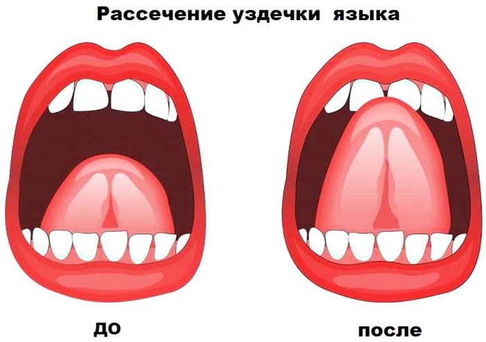
The presence of an overdeveloped frenum is characterized by the appearance of a gap between the lateral and central incisors. In this case, the patient is referred for additional diagnostic procedures, with the help of which it will be decided whether it is necessary to dissect it or apply plastic surgery.
In order to examine the buccal cords, the dentist takes the cheek to the side, after which the expression of the tissue structures of the mucous membrane, which are localized near the alveolar process, is determined. The normal level should be characterized by mild strands. To inspect the frenum in the tongue, the patient must lift it, after which the dentist checks the element with a mirror.
A healthy frenum of the tongue has a thin and long structure, while it is woven into the middle of the organ and the cavity of the mouth. Pathological processes can disrupt its functional feature, which gives the patient additional inconvenience and painful sensations.
On the contrary, an excessively short bridle can disrupt the process of sucking, swallowing, or significantly spoil the quality of speech, especially when pronouncing the sound [p]. Also, such a pathological process can lead to the development of an incorrect bite or periodontal disease.
Periodontal assessment
Examination of the oral cavity (the algorithm of which must be carefully followed by the dentist) includes a value judgment about the functional characteristics of the periodontium. This allows you to more effectively determine the pathological condition or take the necessary preventive measures.
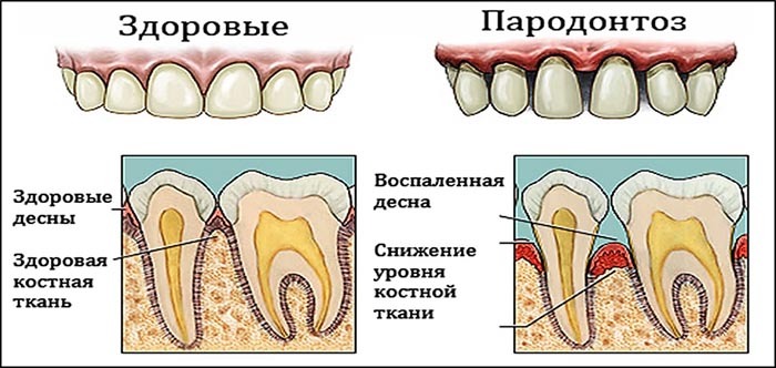
In normal form, the papillae of the gums are well pronounced, pinkish in color and triangular in shape. They also adhere quite tightly to the surface of the teeth, while completely filling the interdental gaps.
A healthy periodontal condition is not characterized by bleeding, regardless of the type of exposure. The natural groove at the gum has a depth of 0.5 mm for the anterior teeth and 3.2-3.5 mm for the posterior ones.
Various deviations from the normative indicator, including severe swelling, hyperemia and bleeding, indicate the presence of a pathological process in the oral cavity. This feature is being investigated in detail by specialists.
Assessment of the condition of the bite
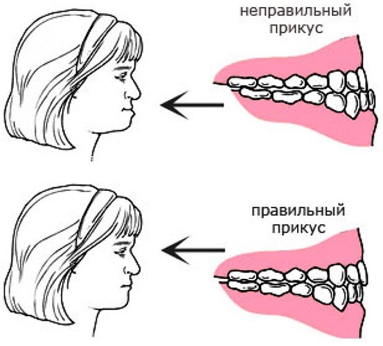
In order to correctly assess the physiological state of the occlusion, it is necessary to determine it based on 3 main positions: the shape of the dental arch, the location of individual elements in the oral cavity, as well as the ratio of the upper and lower jaw.
The main research work is carried out due to the precise fixation of the jaw during swallowing, which allows you to visually determine the nature of clinical manifestations.
Symptomatic manifestations of bite include the following features:
| Localization in the plane | Peculiarities |
| Sagittal | A mesial tubercle located in the upper part of the jaw apparatus; the canines are on either side, with the lower one slightly closer to the center. |
| Vertical | Fissure-tuberous contact is observed between several antagonists in the oral cavity; the overlap of the incisor type is no more than 50% of the height of the tooth crown. |
| Horizontal | The buccal tubercles are located in relation to the lower molars, as indicated by the presence of a central generatrix between them. |
Any clinical judgment should be made with open jaws. In this case, the characteristic signs appear, which are described in the table above. The dentist should pay special attention to the presence of pathological processes, including erosive formations.
A natural bite is possible only when each of the teeth occupies a position characteristic of a certain numerical value. The absence or presence of additional elements represents a degenerative change in the functionality of the bite itself.
Assessment and registration of the condition of the teeth.
In the course of clinical research, the doctor assesses the general condition of the dental crowns, tissues, as well as the exposed part of the root system. For a complete analysis of the outer side of the tooth, it is pre-dried, after which tactile and visual diagnostics are performed.
Such events provide data on the following indicators:
- type and shape of the crown on the tooth, the norm of which is determined in relation to the anatomically correct position;
- enamel surface quality, which should have a holistic structure, uniform density, light tones and shine;
- the presence of restored teeth, including the quality of the operation. Here, the doctor pays attention to the presence of orthodontic structures in the oral cavity, regardless of whether they are removable or not.
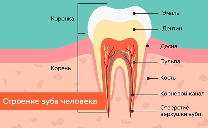
A comprehensive study includes a detailed analysis of the visible side of the tooth base, including medial, oral, distal characteristics. During diagnostic procedures for molars and premolars, the doctor notes the occlusal features of the tooth.
In order not to miss important details, the dentist follows a specific sequence of actions that allow you to examine the surface of the teeth more effectively. So, the research activity begins with the upper right element, after which each tooth is analyzed in turn. The inspection ends with the last element on the left side.
Due to the fact that in dental practice, conventions are used in relation to each tooth and its condition, this greatly facilitates the recording of the condition the patient.
So, the dentition is mentally divided into 4 separate quadrants, where each is assigned its own number - from 1 to 4 for a permanent bite and from 5 to 8 for a temporary diagnosis.
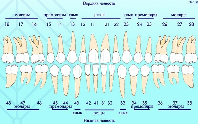
The name of each of the identified teeth, which must be described, is made up of 2 integers, where the first number defines the quadrant, and the second is the number of the tooth itself. For canines, incisors and molars, the conventions apply, which are an exception to the above rule.
Such a dental research methodology allows faster and more effectively investigate, as well as register various pathological changes in the bone dental tissue person.
The main pathological changes affecting not only the teeth, but also the oral cavity can be identified with the help of a preliminary examination. Diagnostics, characterized by a detailed algorithm, makes it possible to start effective treatment in a timely manner, as well as to avoid dangerous complications in the future.
Oral examination video
How is the examination of the oral cavity by the dentist: