
Content
- Characteristic
- Functions and properties
- Anatomy and structure
- Possible diseases and pathologies
- Otosclerosis
- Hyperacusis
- Congenital absence of the oval window
- Tympanosclerosis
- Perilymphatic fistula
- Complications of acute otitis media
- Inner ear barotrauma
- Video about the structure of the ear
The auditory system is relatively simple compared to other senses. This is due to the fact that the process of converting sound vibrations into nerve impulses is linear. Sound is transmitted from the ear to the auditory nerve, and from it to the brain through a chain of internal structures, one of which is called the oval window.
Characteristic
Capturing vibrations by a living creature requires differentiated receptors in it. In the case of sound vibrations, these receptors are located in the inner ear, and their activation requires a preliminary vibration stimulus, which is carried out in the outer and middle ear.
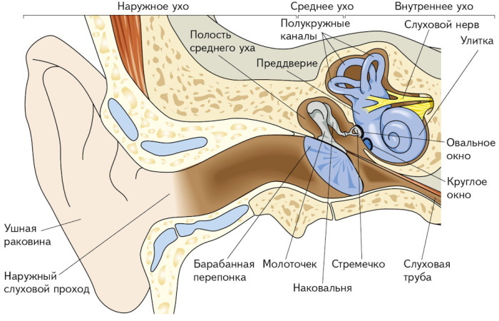
Sound waves entering the auricle pass through the ear canal ≈2.5 cm long and ≈7 mm thick to a thin membrane. This is the eardrum that separates the outer ear from the middle ear. In its cavity there are 3 auditory ossicles: a hammer, an anvil and a stapes. The hammer-shaped bone rests on the eardrum with the handle. The head of the hammer controls the anvil, which transmits the movement of the stirrup, and it rests with its foot plate on the oval window.
The oval window in the ear is a membrane-covered opening that connects the middle of the hearing aid to the inside of the ear. Sound waves vibrate the eardrum, and the bones transmit vibrations to the oval window, which causes fluid to move inside the cochlea and activates hearing receptors.
Functions and properties
The middle part of the ear is located between the outer and inner parts of the organ.It is necessary for a smooth transition of the incoming sound stream to the structured sound. The function of the outer ear is to pick up sound vibrations and direct them to the inner parts. In this process, some of the received audio frequencies increase while others decrease.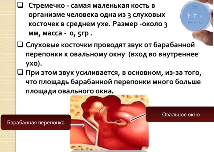
If the sound goes directly from the external passage to the liquid (perilymph and endolymph) located in the inner ear, then large losses when a sound wave is reflected, since the resistance of an acoustic wave to an aqueous medium is several orders of magnitude higher than the resistance air. Therefore, it is necessary to adapt the acoustic impedances, which is what happens within the median part of the ear instrument.
The middle ear transmits the movement of a sound wave from the eardrum to the inner ear. There is an increase in pressure on the connective tissue of the oval window, which is transmitted through the stapes. As a result, the sound wave presses on the cochlea. When waves are transmitted from the eardrum to the oval window, the middle ear functions as acoustic transducer, amplifying sound waves before they reach the inside hearing aid.
The pressure of sound waves in the oval window is about 20 times higher than in the eardrum. It increases due to the difference in size between the relatively large surface of the tympanic membrane and the small surface of the oval window. The same principle applies when a person steps on the foot in stiletto heels. A small surface is much more painful than a flat shoe with a large surface area.
When the vibrations of the auditory bones are transmitted to the perilymph in the inner ear, there is almost no loss of sound absorption. The eardrum and the ossicle increase sound pressure because it is transmitted from a large area (eardrum) to a small area (oval window) in a 17: 1 ratio.
Together with the lever of the sound-conducting chain (hammer, anvil, stirrup), the pressure on the oval window lined with a membrane increases 22 times. Further transmission of pressure fluctuations occurs from the stapes to the practically incompressible perilymph, which makes forced vibrations. From there, the sound travels through the auditory nerve to the brain, which processes it.
The oval window in the ear can be injured by excessive sound pressure and then its membrane will be broken. The fluid from the inside (perilymph) will seep into the middle ear, forming a perilymphal fistula. Fluid transfusion leads to damage to the organ of hearing, and sometimes the vestibular apparatus.
Anatomy and structure
The middle section is an air-filled cavity that communicates with the air cells of the mastoid process of the temporal bone and the antrum of the mastoid process by means of aditus (narrow isthmus). The eustachian tube, about 1 mm wide and 35 mm long, connects the middle ear to the nasopharynx, and its function is to balance the pressure of the middle ear with the pressure of the atmosphere. The structures of the tympanic cavity are covered by the mucous membrane.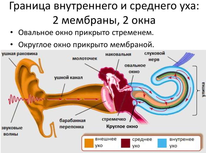
The epitympanic depression (notch located at the top of the middle ear) contains the head of the malleus, the body, and the short stem of the incus. The hammer represents the head and neck. The anvil consists of a body, a short and a long stem. The stapes is the head, anterior and posterior legs, and the base or plate of the foot, which is attached by an annular ligament to the edge of the oval window. They are held in place and moved by joints, muscles and ligaments that help transmit sound.
The bones of the middle ear amplify sound waves and send them to the cochlea. It is a structure in the form of a spiral, the channels of which are filled with liquid. Located in the inner ear. The top and bottom of the cochlea are separated by an elastic “basilar” membrane that serves as the base or ground floor on which the key hearing structures are located.
The incoming sound waves cause the fluid inside the snail to vibrate. Under the influence of air movement, a traveling wave is formed along the basilar membrane. Together with the wave, the hair cells that are on the membrane move up and down.
The bristly hairs of the cells collide with the overlying membrane, as a result of which the bristles tilt to one side and open the porous channels. Certain chemicals are then released, creating an electrical signal that travels through the auditory nerves to the brain. The end result of these manipulations is a recognizable sound.
The hair cells (receptors in the auditory system) at the base of the cochlea pick up high-frequency sounds, such as the ringing of a cell phone. Those closer to the middle pick up lower sounds, such as the barking of a large dog.
In the ear, behind the stapes, there are 2 small holes through which the cochlea connects to the tympanic cavity. It is an oval and round window. With the help of the first, the cochlea communicates with the middle ear cavity. It is closed by the foot plate of the stapes. The round window provides an outlet for sound vibrations.
The blood supply to the middle part of the ear apparatus comes mainly from the maxillary part of the external carotid artery, which is supplemented by the branches of the internal carotid artery. The deep ear artery supplies blood to the tympanic cavity and also supplies the outer side of the eardrum.
The middle ear is innervated by the ear-temporal (V cranial) and tympanic (IX) nerve endings and the auricular branch of the vagus nerve.
Possible diseases and pathologies
Hearing impairment is not an ear disease, but a consequence that always needs to be clarified with an ENT doctor.
Violations are divided into 3 main types:
| Conductive hearing loss | Sound waves reach the inner ear less or not at all. The cause may be a malformation or blockage of the ear canal. Conductive hearing loss in the middle ear is often the result of an infection. Victims perceive everyday noises much more quietly and hear as “through cotton wool”. |
| Sensorineural hearing loss | Most often affects the inner ear. Sound waves travel from the eardrum and ossicles to the inner ear, but are not properly processed and do not travel to the hair cells. If the damage is caused by prolonged exposure to noise, it is said to be noise-induced hearing loss. |
| Hearing loss in the elderly (presbycusis) | The most common form of sensorineural hearing loss. It occurs as a consequence of the body's natural degeneration processes in old age. |
Window anomalies are defects in the bone structure of the inner ear that lead to hearing loss. Normal sound transmission is transmitted through oval and circular windows, which serve as contacts between the air in the middle ear and the fluid spaces of the inner ear. Various conditions can enlarge bone canals or create additional defects in the bone labyrinth. All disorders of the middle ear lead to hearing impairments.
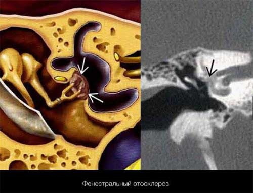
Otosclerosis
Due to hardening and new bone formation on the stirrup, hammer and incus, the bones can no longer adequately transmit sound. The consequences are hearing loss and possibly tinnitus. The reasons are not fully understood, but, according to doctors, there are links with viral infections, impaired autoimmune processes and hereditary predisposition.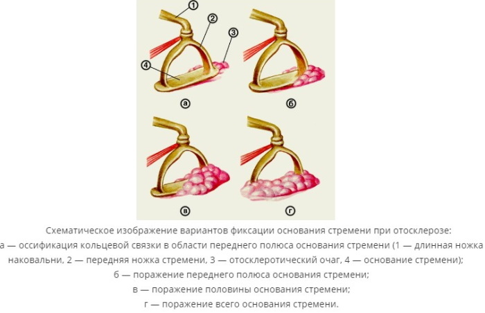
The disease is characterized by abnormal bone growth in the middle ear region. Usually, the disorder affects the stapes, a bone in the area of the oval window. It is in this window that the stirrup base plate comes into contact with the fluids of the inner ear and acts as piston, conducting sound energy from the tympanic membrane into the labyrinths of the inner part surrounded by perilymph.
In otosclerosis, the gradual build-up of new cancellous bone around the stapes welds it to the wall and immobilizes it, preventing vibration and preventing sound waves from passing through the ear. The result is hearing loss. In the later stages of the disease, when otosclerosis progresses and affects the structures of the cochlea, sensorineural hearing loss develops, affecting the inner ear. It often develops in conjunction with conductive hearing loss.
The process leads to progressive hearing loss because the bone's ability to vibrate becomes increasingly limited.
Clinical signs of the disease:
- Slowly progressive hearing loss.
- The defeat of the second ear in 70% of patients as the disease progresses.
- Tinnitus
- Slight dizziness.
- Quiet speech.
- Schwarze's symptom develops: a red-blue spot appears in the ear canal, visible through the eardrum.
It is believed that many cases of otosclerosis are inherited. Middle-aged women are at greatest risk.
Hyperacusis
Hyperacusis is a relatively rare hearing disorder whose main symptom is an increase in everyday sounds. While hyperacusis is not dangerous, it can make life difficult, exacerbate social relationships, make work difficult, and add unnecessary stress.
In the case when the stapes bone is paralyzed, there is an incorrect perception of sounds. Weak waves are perceived as overly intense. The foot plate of the stapes moves away from the oval window, which makes it impossible to reduce vibration, so a constant noise is heard in the ear.
Congenital absence of the oval window
The oval window in the ear may be missing due to a birth defect. A rare embryological defect is characterized by manifestations of conductive hearing loss from birth or early childhood.
Most children with conductive hearing loss are eventually diagnosed with acquired otitis media or middle ear effusion. The absence of the oval window is uncommon and may be due to an abnormality of the horizontal portion of the facial nerve canal. Congenital absence of the oval window is diagnosed on computed tomography.
Tympanosclerosis
This is a condition in which the middle ear is affected, causing the eardrum to become bright white. The whiteness is due to calcium deposits. In addition to the membrane, the oval, round window and bones can be affected.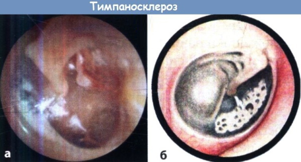
The most common symptom of tympanosclerosis is conductive hearing loss. The only treatment is surgery to repair the eardrum and other involved structures of the middle ear. Calcination promotes immobilization of the stapes, without the action of which it is impossible to create sound. In these cases, plastic or prosthetics of the stapes are performed. If the treatment was done late, then hearing aids are one of the most effective solutions for hearing restoration.
Perilymphatic fistula
The oval window in the ear may not function due to a defect in the cochlear labyrinth, when the ratio of the perlimph in the cochlea is disturbed, and as a result, a fistula develops. Most often, fistula occurs after traumatic brain injury, barotrauma, iatrogenic injury. An internal fistula of the tympanic membrane disrupts the normal hemodynamics of the inner ear. This leads to vestibular and auditory disorders.
Symptoms of a perilymphic fistula include dizziness, imbalance, nausea, and vomiting. Some people experience ringing (tinnitus) and a stuffy feeling in their ears, and many people experience short-term hearing loss. Symptoms worsen with changes in altitude (elevator, plane, or mountain travel), weather, and exercise.
If the fistula is small, then this condition is treated conservatively with bed rest in a sitting position. Stress, sneezing, blowing your nose, sexual activity, loud noises should be avoided to prevent pressure waves in the inner ear. A fistula in the area of a rounded window often heals within a week or two with this regimen.
Medium-sized fistula lends itself to conservative treatment, but if the diameter of the fistula reaches a size of more than 2 mm, and hearing loss progresses or other signs persist, surgery may be required intervention.
Complications of acute otitis media
Inflammation of the middle ear cavity often leads to damage to the facial nerve and labyrinth. This is due to various reasons. Distinguish between hematogenous, meningogenic, traumatic and tympanogenic labyrinthitis. The latter develop as complications of acute and chronic forms of otitis media. In acute otitis media, there is a change in the permeability of the annular ligament of the oval window, the membrane of the round window, as a result of which bacterial toxins penetrate into the inner ear.
Inner ear barotrauma
A malfunctioning device creates negative pressure in the middle ear during submersion. To compensate, the eardrum bulges inward and deflects the oval window towards the inner ear through the ossicular chain. If the diver tries to forcibly equalize the pressure, then it increases in the inner part through the channels of the cochlea.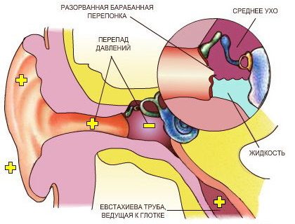
This increase in pressure, combined with the deflection of inner ear structures due to the inwardly curved eardrum, can rupture the circular window membrane. The perilymph will flow from the inner to the middle, and the nourishment of the inner ear structures will be disrupted or suspended.
If function is impaired, excess pressure can build up in the middle ear, causing the eardrum to protrude into the ear canal. The oval window can be bent outward, and the membrane of the round window inward with the help of the auditory ossicles. In the event of a sudden equalization of pressure, mechanical damage to the inner ear or damage to the membrane of the round window can occur.
There are different treatment options:
- Cortisone therapy.
- If necessary, operation with sealing the membrane of the round window.
- Training of the vestibular apparatus.
An oval or round window plug is a stabilizing procedure sometimes used to repair perilymphal fistulas to stop fluid leakage. Middle ear surgery is performed using a transcanal endoscopic approach. The holes in the oval or round window are closed with tissue taken from the outside of the organ or behind the ear so that perilymph fluid does not seep through the fistulas.
This operation requires a long postoperative period, followed by activity limitation to ensure the success of the graft. Possible adverse outcomes include potential hearing loss and worsening symptoms.
It is impossible to pinpoint when a hearing loss will occur - it is usually only recognized when it is too late. Hearing loss occurs when constant noise damages the inner ear, its structures: bones, oval window, membranes.
In many cases, the disease develops gradually. Therefore, it is recommended to be checked regularly. This will allow the first warning signs to be recognized and appropriate treatment initiated. Hearing impairment and hearing loss impair quality of life, affect relationships between people, and can lead to health problems such as dementia, cardiovascular disease and depression.
Author: Belyaeva Anna
Video about the structure of the ear
Anatomy and physiology of the organ of hearing:


