
Content
- Characteristic
- Functions and properties
- Anatomy and structure
- Possible diseases and pathologies
- Hyporeflexia
- Hyperreflexia
- Thyroid dysfunction
- Adi syndrome
- Diabetes
- Achilles reflex video
Achilles test reflex - simple and effective part of a complete neurological examination of the lower extremities. The deep tendon response provides insight into the function of the reflex arc, whether it is symmetrical or not.
Characteristic
The reflex arc is a neural pathway in the human body that connects muscle groups without affecting the brain. Pathways control involuntary movements in response to a specific stimulus. One example is the rapid blinking of the eyes in response to dust in the air, coughing when food gets stuck in the windpipe, and a kick in the center of the knee. Reflex arcs are completely independent of the paths along which most nerve impulses travel. But the messages they convey are just as important, which is why impaired reflexes often signal serious problems with nerve control and muscle support.
The arches are usually located in the spine or brain, although there is no conscious control in either of these locations. Reflex arcs are shorter than longer neural pathways. Those in the spine control the responses of the large muscles in the arms and legs, while those in the brain are associated with responses in the face.
The doctor palpates the Achilles tendon just above the back of the calcaneus. It is the strongest and largest tendon in the human body and attaches the superior triceps muscle (soleus and gastrocnemius) to the calcaneus tubercle. The tendon is covered with paratenon, a thin sheath that reduces friction while also serving as a conduit for blood vessels.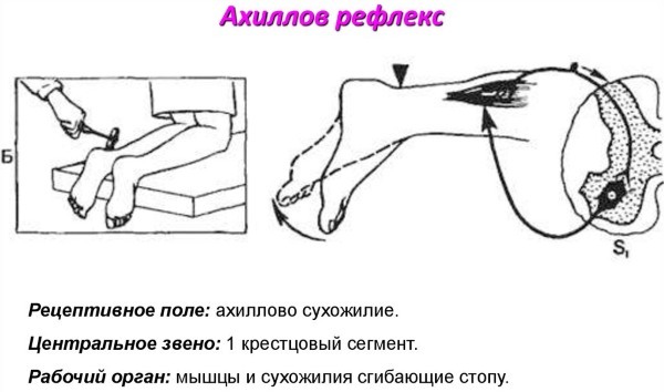
The Achilles reflex (the reflex arch consists of sensory and motor fibers of the tibial nerve) is called an ankle reflex test. This is a deep tendon response that can be easily triggered with a standard instrument, the reflex hammer. The Achilles Tendon Test is a simple and effective part of a complete lower limb examination and physical nervous system screening.
Functions and properties
The test is performed as part of a complete physical examination to assess the neurological function of the lower extremities. When the question arises about the pathology of the S1 nerve root, the Achilles reflex test is the initial assessment of choice. This helps to differentiate the pathology of the UMN (component of the upper motor neuron) from the pathology of the LMN (component of the lower motor neuron), and also helps to determine the location of the lesion.
In addition, the reflex arc response can provide information about peripheral nerve function. This is sometimes of paramount importance for certain patient groups, such as those with diabetes and hypothyroidism.
The ankle reflex is also called the Achilles reflex. The doctor taps the tendon with the foot toward the legs with the toes toward the lower leg. The position is called bent back. The normal response should be a downward jerk of the foot due to the calf and soleus reflex. When there is no ankle response, diagnostic measures are initiated.
The Achilles reflex (the reflex arc consists of receptors that perceive irritation) is also detected in response to a blow to the heel tendon. This alternative method for assessing the tendon reflex is useful for hospitalized patients with limited mobility.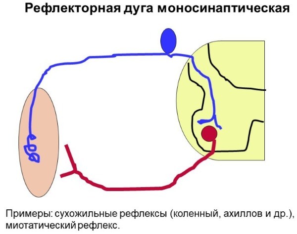
It is performed by tapping the forefoot of the patient with the toes. This triggers flexion of the foot or the plantar flexion reflex. This raised leg method has been shown to provide the most reliable results in older patients.
Anatomy and structure
The Achilles tendon, or calcaneus, is a tough strip of fibrous tissue that connects the calf muscles to the heel bone. The calf and soleus muscles combine into one strip of tissue that becomes the Achilles tendon at the lower end of the lower leg. It is then inserted into the heel bone.
The heel tendon is the largest and strongest connective tissue formation in the body. When the calf muscles flex, bundles of collagen fibers are pulled at the heel. This movement allows you to lean on your toes when walking, running, or jumping. Despite their strength, the fibers are vulnerable to injury due to the limited blood supply and high stress they experience.
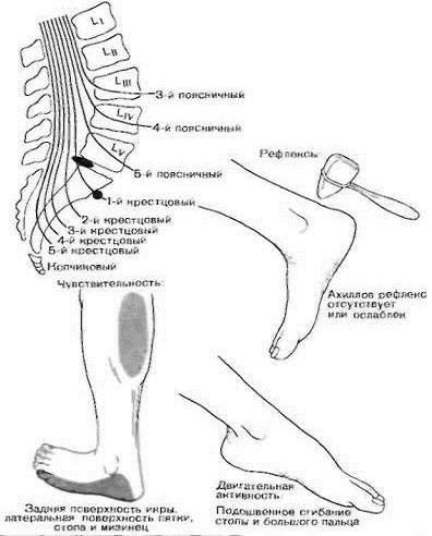
The Achilles tendon is innervated by the S1 and S2 nerve roots of the tibial nerve. The tendon reflex is a stretching response that refers to the involuntary contraction of a muscle in response to passive stretching.
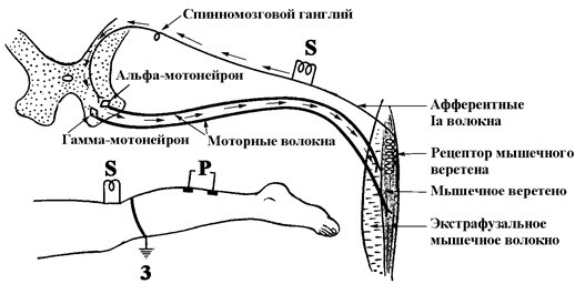
When tapping the tendon with a reflex hammer, the triceps muscle is lengthened (stretched) and the following chain of actions occurs:
- Stretch receptors in the muscle spindle are activated and excitatory stimuli are propagated.
- The stimulus is carried by afferent neurons (Ia) to the spinal cord.
- Afferent neurons synapse and activate an alpha motor neuron in the ventral horn of the spinal cord.
- The activated alpha motor neuron transmits the efferent stimulus back to the neuromuscular junction of the agonist muscle (triceps), resulting in muscle contraction (reflex).
To ensure that the muscle agonist (triceps surae) of the reflex has an unobstructed contraction, afferent neurons Ia simultaneously stimulate an inhibitory neuron in the spinal cord that prevents contraction antagonist muscles.
The spinal reflex arcs bypass the brain to provide a quick response.
But the descending corticospinal pathways from the cerebral cortex weaken and reduce the amplitude of the reflex arc. The S1 nerve root serves as a conductor of both afferent (responding to various stimuli) and efferent (transmitting signals) impulses of the reflex arc of the Achilles tendon.
The deep tendon reflex arc is divided into an upper motor neuron (UMN) component and a lower motor neuron (LMN) component. The UMN component consists of descending structures: the corticospinal tract, brainstem, internal capsule, and cerebral cortex. LMN structures are the ventral (anterior) horn of the spinal cord and the peripheral nerve that innervates the muscle. The LMN of the Achilles reflex consists of the ventral horn of the S1 nerve root and the tibial nerve.
Possible diseases and pathologies
The Achilles reflex (the reflex arc does not directly affect the brain) is interpreted and evaluated on the basis of the myotic reflex scale.
It uses a grading system from 0 to 4:
| 0 | There is no reflex. |
| 1 | The reflex is weak, less than normal. |
| 2 | The reflex is in the lower half of the normal range. |
| 3 | The reflex is in the upper half of the normal range. |
| 4 | The reflex is enhanced, more than usual, including clonus if present. |
In addition, a plus (+) or minus (-) can be added to the score to indicate an intermediate stage. A score of 5 can also be used to indicate a sustained clonus of> 10 rhythmic oscillations after rapid muscle stretching.
Reflexes can also be diagnosed in a descriptive way:
- absent;
- hypoactive;
- normal;
- hyperactive.
Testing deep tendon reflexes gives the doctor information about the state of the motor nerves, the forms of synaptic connections in the spinal cord, and descending motor conductors. Damage to the upper motor neuron, UMN, causes an increased reflex, while damage to the lower motor neuron, LMN, causes hypoactivity of the reflexes. In the study, any asymmetric increase or decrease in reflexes is noted.
Lack of ankle reaction can be associated with over 47 different diagnoses.

Some of them:
- Poisoning with drugs or alcohol.
- Familial genetic disorders.
- Spina bifida.
- Vitamin E deficiency.
- Injury, such as concussion.
When examining deep tendon reflexes, it is extremely important to compare each reflex response with the corresponding opposite side. Hyporeflexia, hyperreflexia, and asymmetric responses are abnormal and require further evaluation.
Hyporeflexia
A decreased muscle reflex usually indicates an LMN disorder. LMN disease or disruption causes reduced or absent reflex because no stimulus is transmitted to the effector muscle. Additional signs of the disease are lethargy or decreased muscle tone, weakness, twitching, and muscle atrophy.
Causes of LMN disease include:
- Acquired peripheral neuropathies.
- Physiological disorders caused by diabetes mellitus, hypothyroidism, uremia, vitamin and electrolyte deficiencies, and toxins such as lead or arsenic.
- Medicines such as isoniazid, vincristine, and diphenylhydantoin are known to cause hyporeflexia.
- There are many hereditary and immune-related causes: atrophy, spinal muscular atrophy, distal hereditary motor neuropathies and syndrome Guillain-Barre.
- Structural damage to the LMN components of the tibial nerve can occur with stenosis of the nerve openings or herniation of the intervertebral disc at the L5-S1 level, which compresses the S1 nerve root.
Hyperreflexia
An increased muscle reflex indicates diseases caused by the pathology of the UMN. Damage to any component of the motor neuron (corticospinal tracts, brainstem, internal capsule, and cortex brain) leads to an overactive deep tendon reflex due to disinhibition of the reflex arc spine.
As previously described, upper motor neurons play an important role in suppressing reflex responses. In addition to hyperreflexia, UMN lesions are manifested by spasticity or increased muscle tone, weakness, and various pathological reflexes.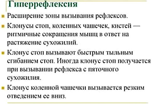
The reasons for damage to the UMN can be caused by:
- anoxic brain damage (encephalopathy);
- trauma to the brain or spinal cord;
- cerebrovascular (cerebrovascular pathology) disorders;
- neurodegenerative disorders (amyotrophic lateral sclerosis, primary lateral sclerosis, multiple sclerosis);
- spinal cord compression;
- malignant neoplasms.
The reflex response of the nerve arc at the time of the Achilles reflex test is used in some cases to identify various syndromes and disorders.
Thyroid dysfunction
Among the many methods developed to study thyroid function, two are considered that investigate peripheral aspects: Achilles bone-tendon reflexion (timing of contraction and relaxation of the gastrocnemius muscle) and the reaction of the cardiovascular system to thyroid hormones glands.
People with hypothyroidism (decreased function) relax slowly. Water, sodium and calcium are excreted more slowly in hypothyroidism. There is also a slow relaxation of the heart muscle, which contributes to heart failure, because the heart in this case cannot receive as much blood as a relaxed heart normally does. The blood vessels in hypothyroidism cannot relax properly, which contributes to hypertension.
Adi syndrome
A neurological disorder that affects the pupil of the eye and the autonomic nervous system. This affects the reflexes of both the pupil and the deep tendons. The larger than normal pupil of one eye tapers slowly in bright light. This occurs along with a lack of deep tendon reflexes, usually in the Achilles tendon. The disease begins gradually in one eye and often progresses to the other eye.
This may initially cause loss of deep tendon reflexes on only one side of the body, but then progresses to the other side. Eye and reflex symptoms may not appear at the same time. People with Adi syndrome may also sweat excessively, sometimes only on one side of the body.
The combination of these three symptoms - abnormal pupil size, loss of deep tendon reflexes, and excessive sweating - is commonly referred to as a variant of Adi syndrome. Some patients may develop cardiovascular problems. The condition appears on its own or in combination with other diseases of the nervous system, such as Sjogren's syndrome or migraine.
Diabetes
In the case of long-term diabetes mellitus, diabetic polyneuropathy develops. It is a debilitating condition that affects nearly 50% of diabetic patients. The main types of diabetic neuropathy are sensorimotor and autonomic.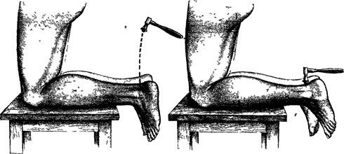
When sensory nerves are damaged, the nerves with the longest axons are affected first. Evaluation of sensorimotor neuropathy reveals loss of reflexes to the Achilles tendon and patella, gait ataxia (impaired muscle coherence), and balance problems.
The Achilles tendon reflex is a simple technique for detecting the reflex arc response, which is part of any neurological examination of the lower extremities. It can be performed both in a clinic and in a hospital setting. However, differences in technique and levels of medical professional experience can lead to different results.
Achilles reflex video
Achilles reflex:


