When reading an electrocardiogram, you can often encounter a rise (significant or not so) ST segment on the ECG waveform. What this sign means, and how it should be interpreted to the patient, cardiologists explain in detail during the examination period.
Record content:
- 1 What is the ST segment and what is it responsible for?
- 2 ST segment measurement rules
-
3 Causes of ST segment elevation
- 3.1 Early repolarization syndrome
- 3.2 Prinzmetal's angina
- 3.3 ST-segment elevation myocardial infarction.
- 3.4 Heart aneurysm
- 3.5 Acute pericarditis
- 3.6 Brugada syndrome
- 3.7 Non-infarct myocardial injury (myocarditis, LV tumor, traumatic ventricular injury)
- 3.8 Hyperkalemia
- 3.9 Systemic hypothermia (hypothermia)
- 3.10 Taking medications
-
4 Clinical manifestations
- 4.1 Pain
- 4.2 Dyspnea
- 4.3 Fever (increased body temperature)
- 4.4 Excessive sweating
- 4.5 Heart rhythm disorders
- 4.6 Weakness
- 4.7 Asymptomatic course
- 5 ST segment elevation video
What is the ST segment and what is it responsible for?
Any electrocardiogram contains teeth, intervals and segments, each of which reflects the propagation of an excitation wave in the heart at one stage or another.
A segment is a section of an electrocardiogram located between two teeth (that is, a segment starts where one tooth ends and ends at the beginning of the next tooth). Accordingly, the ST segment should be considered as the segment between the S and T waves.
This segment corresponds to a state of the heart muscle in which both ventricles are at the stage of returning to rest. It cannot be said that this is already a complete relaxation of the myocardium, just as it cannot be called a contraction process.
The medical term that most accurately characterizes this condition of the muscular wall of the heart is called repolarization in the scientific medical literature. The QRS complex corresponds to the contraction of the ventricles, the T wave corresponds to relaxation. The ST segment clearly demonstrates the transition from the state of contraction of the heart muscle to its relaxation.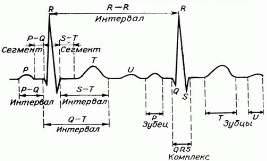
Depending on the analyzed lead, it is possible to evaluate the process of excitation of one or another part of the heart muscle:
| Lead ECG data | What characterize |
| V1 and V2 | They give an idea of the processes taking place in the interventricular septum. |
| V3 and V4 | Inform about the state of the anterior myocardial wall. |
ST segment measurement rules
In order to eliminate errors in assessing ST segment indicators, it is necessary to adhere to a number of generally accepted rules:
- First, you need to find the junction point on the ECG (also called the J point), which is the end of the S wave. If the S wave is absent, then this point is the point of intersection of the descending knee of the R wave with the isoelectric line (the horizontal line between the T and P waves).
- Next, you need to retreat by 0.08 seconds (at a tape speed of 50 mm / s, this distance corresponds to 4 mm of film, at 25 mm / s - 2 mm), and from here start measuring the segment under study.
The rise of the ST segment on the ECG can be diagnosed correctly only if these rules are observed, otherwise you can make a mistake and skip the rise of the initial part of this segment (the so-called Osborne wave), which is of particular importance in diagnosing a series diseases.
When evaluating the ST segment on an ECG, the following significant parameters are taken into account:
- The duration of the segment (its length, measured in seconds).
- The deviation of the connection point from the isoline (it can be positive (+) if the point is shifted up, or negative (-) if the point is shifted down);
- The position of the ST segment relative to the isoelectric line (the segment can be located on the isoline, its depression (downward deviation) or elevation (rise) can be observed.
Normal values for adults
Normally, in the adult population, the ST segment coincides with the isoelectric line (there may be a deviation of 0.5 mm up or down from it). For some leads, a larger offset is acceptable.
So, in the chest leads V1-V3, a displacement of up to 2 mm upward is possible, provided that the T wave is positive. Elevation of the ST segment in other ECG leads is considered abnormal. In leads V4, V5, V6, a segment depression of up to 0.5 mm is possible.
What is the ECG with a significant ST elevation talking about?
If the analysis of the electrocardiogram revealed an elevation of the ST segment of more than 0.5 mm (or more than 2 mm in V1-V3), we can talk about a pathological elevation of the ST segment on the ECG.
In most cases, its root cause is ischemic damage to the heart muscle. Ischemia is a decrease in blood flow to an organ or tissue that can impair the function of the affected area.
The human body, thanks to various compensation mechanisms, has the ability to function under ischemic conditions without organic disturbances (that is, there are no violations of the integrity of an organ or tissue).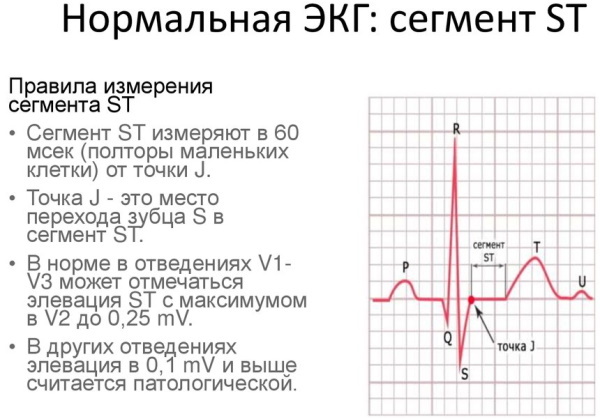
However, with prolonged ischemia, when the affected area is in deficiency conditions for a long time blood supply, there is a depletion of compensation systems, as a result of which, in turn, there is already organic damage. In the early stages, it is still reversible.
If damage affects the entire thickness of the myocardium (such damage is called transmural), or is localized under the epicardium (the part of the heart wall located above the myocardium), then on the ECG there will be a rise in the segment ST.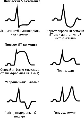
In transmural injury, the elevated ST segment often fuses with the T wave, forming a characteristic curve. Such a fusion is called in medical terminology "cat's back" or "death wing".
Causes of ST segment elevation
Ischemic damage can be observed as a result of various clinical conditions, which will be discussed in detail below.
Early repolarization syndrome
Most authors consider the presence of this syndrome to be a variant of the norm. This phenomenon may not manifest itself clinically in any way and is established only by ECG results. The elevation of the ST segment along with a change in the electrical axis of the heart (it rotates counterclockwise) are the main signs of this syndrome.
Prinzmetal's angina
Angina pectoris consists of an attack of pain behind the breastbone. This symptom is typical for patients with coronary heart disease. It usually occurs in response to emotional stress during physical exertion.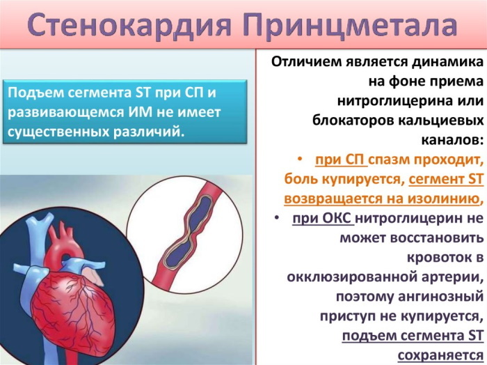
However, in about 25% of cases in the observed patients, the pain syndrome manifested itself at rest. Pain with angina pectoris is short-lived, and is stopped by taking nitrates, which distinguishes angina pectoris from myocardial infarction.
If, during an attack of angina pectoris, a cardiogram is made, then a change in the T wave will be observed, as well as a displacement of the ST segment below the isoline.
Elevation of the ST segment, together with the absence of other changes recorded on the ECG during an attack, is a specific sign of Prinzmetal's angina. This is a rare form of angina pectoris. It has no other electrocardiographic signs, but to make a diagnosis, it is imperative to reconcile the ECG data with clinical manifestations in the anamnesis.
ST-segment elevation myocardial infarction.
Heart attacks are the most common diseases accompanied by an elevation of the segment in question. Myocardial infarction is a pathological condition based on necrosis (death) of a portion of the heart muscle.
The cause of a heart attack is most often a violation of the blood supply to the heart as a result of the overlap of the lumen of the vessel by an atherosclerotic plaque, thrombus, rupture of the vessel wall and a number of other reasons.
The rise of the ST segment on the ECG is not a specific sign of a heart attack and will be observed only in the stage of damage (this sign remains intact from several hours to several days).
Depending on the stage of development of the pathological process, other signs will be observed, including a change in the T wave, as well as the appearance of a pathological Q wave. There is also a form of myocardial infarction that is not accompanied by ST segment elevation.
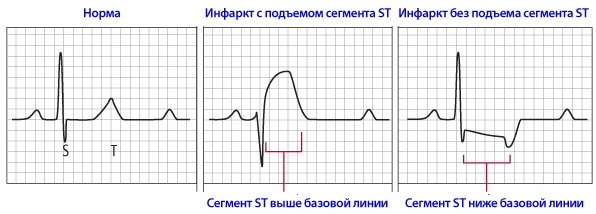
In acute myocardial infarction, the ST segment elevation can be of different forms: in the form of a dome, a plateau, an oblique form. It should not be forgotten that ST segment depression will be observed in the reciprocal (mirror) leads.
This is due to the peculiarities of the placement of electrodes and the propagation of excitation during ECG registration. To diagnose myocardial infarction at the initial stage of diagnosis, clinical symptoms and ECG changes are quite enough.
Heart aneurysm
An aneurysm is an abnormal swelling of a portion of the heart wall. During the work of the heart, during contraction, there is an even greater protrusion of the aneurysm outward. As a rule, such bulging is often formed due to myocardial infarction, on days 20-21, at the site of muscle fiber necrosis. Aneurysm can be both acute and chronic.
The above changes in the cardiogram with aneurysm are associated with the presence of damage during myocardial infarction. A number of authors believe that during the formation of the aneurysm, the damage is still ongoing, therefore, there is elevation.
A distinctive feature of the electrocardiogram in this pathological condition is that the ST segment remains permanently elevated, and other changes in the electrocardiogram characteristic of an aneurysm also do not change over time, the ECG takes the form "Frozen" curve. However, the absence of a persistent rise in the ST segment on the ECG does not at all exclude the presence of this pathology.
Acute pericarditis
Pericarditis is inflammation of the pericardium (the outer lining of the heart). It can arise as a result of various infectious diseases, tumors, systemic connective tissue diseases, surgical interventions and more. ECG signs of pericarditis resemble the acute stage of myocardial infarction.
The rise of the ST segment on the ECG of a patient with pericarditis is the earliest sign of this pathology. The criteria for excluding myocardial infarction are the leads in which this picture is observed. With a heart attack, changes affect only those leads that reflect the processes occurring in the affected area of the myocardium.
The pericardium covers the entire heart, therefore, changes in the ECG affect a larger number of leads (up to all with extensive damage). With pericarditis, there is no pathological Q wave.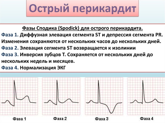
Changes in the PR segment are also characteristic, the appearance of negative T waves is possible.
Brugada syndrome
Severe hereditary pathology, represented by a combination of syncope (fainting) conditions and sudden cardiac death in patients without organic disorders of the cardiovascular systems.
The main electrocardiographic signs of this condition are various types of arrhythmia (ventricular tachycardia, ventricular fibrillation), however, the rise of the ST segment on the ECG with this pathology is also recorded quite often.
Non-infarct myocardial injury (myocarditis, LV tumor, traumatic ventricular injury)
Myocarditis is an inflammatory disease of the muscular layer of the heart. May be caused by exposure to infectious (viruses, bacteria) and non-infectious (eg traumatic) agents.
The electrocardiographic picture of myocarditis is variable and often nonspecific. The rise of the ST segment on the ECG with myocarditis can be found quite often. It can be combined with signs of rhythm and conduction disturbances and even with a pathological Q wave, due to which myocarditis can be confused with myocardial infarction.
Hyperkalemia
Normal serum potassium levels are within the 3.5-5 mmol / L range. An increase in potassium levels of more than 5 mmol / L is dangerous and can be fatal.
Typical electrocardiographic signs of hyperkalemia are:
- Increase in the T wave, its sharpness;
- Elevation of the ST segment;
- Prolongation of the P-R interval, decrease in the amplitude of the T wave.
- Extension
In hypokalemia, the ST segment, on the other hand, is located below the isoline.
Systemic hypothermia (hypothermia)
The peculiarity of the ST segment in this state is that there is a hilly rise at the beginning of the ST complex, that is, in fact, there is an elevation of the J point. This phenomenon is called the Osborne wave. When warmed up, the waves disappear.
Taking medications
When taking cardiac glycosides (for example, Digoxin), a trough-like change in the ST segment is possible (both elevation and depression can occur).
In addition to the above nosologies, the ST segment elevation on the ECG can also be observed with hypertrophy of the left ventricular wall, as well as with blockade of the left bundle branch.
Clinical manifestations
The clinical presentation of ST segment elevation can vary. It depends on the cause of the change.
Pain
The most common symptom accompanying ST segment elevation is pain. The intensity of pain, its duration, irradiation (conduction of pain to other organs and parts of the body) may vary.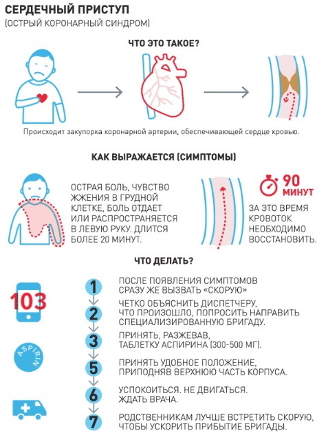
Pain in angina pectoris and myocardial infarction, as a rule, is acute, very severe. It is impossible to endure such pain. By nature, it can be burning, squeezing, pressing.
The localization of pain can also be different. Most often it is localized behind the sternum, directly in the region of the heart. If a heart attack affects the lower wall of the heart, then the abdomen can become the epicenter of pain, which is why patients mistakenly turn to a gastroenterologist.
Often the pain is not localized in one place, but radiates along the course of the nerves. In this case, pain in the heart can be given to the left hand, shoulder blade, shoulder, little finger. There are cases when the only symptom of myocardial infarction was pain in the little finger.
One of the main criteria for differentiating pain in angina pectoris and pain in myocardial infarction is time. Pain with angina pectoris lasts no more than 15 minutes; in all other cases, a heart attack should be suspected.
Pericardial pain is usually similar to the pain of myocardial infarction. It has the same localization, the same areas of conduct. It is distinguished by strengthening in a horizontal position (lying), when eating or drinking (it is especially difficult for patients to swallow), when coughing. Sometimes there is an increase in pain during breathing.
Dyspnea
Shortness of breath is the equivalent of pain in conditions accompanied by an elevation of the ST segment on the ECG. It can occur at rest, during physical exertion. This symptom can manifest itself with myocardial infarction, myocarditis, heart aneurysm.
Fever (increased body temperature)
This nonspecific symptom accompanies many human diseases, and the rise of the ST segment can also manifest itself as an increase in body temperature.
Excessive sweating
It is observed in myocardial infarction, aneurysm and other conditions accompanied by ST segment elevation on the ECG.
Heart rhythm disorders
This symptom can be subjectively felt as increased, or vice versa, weakened heartbeat, interruptions in the work of the heart.
Weakness
General weakness, weakness of skeletal muscles may also indicate the presence of heart disease, accompanied by an elevation of the ST segment.
Asymptomatic course
In some cases, ST elevation may not manifest itself clinically. This situation is especially dangerous, since some conditions accompanied by an elevation of the ST segment on the ECG are often life-threatening and require urgent medical attention.
ST segment elevation video
Elevation of the ST segment: