
X-ray ankle examination - this is one of the most modern and most informative diagnostic methods, which allows you to quickly and accurately determine the cause of the painful condition of the lower limb, make the correct diagnosis and form an effective course therapy.
Ankle X-ray requires complex preparation and is a painless procedure with a minimum of contraindications. The appointment and interpretation of the diagnostic procedure is carried out by the attending physician of the appropriate qualifications.
Record content:
- 1 Indications for research
- 2 How is it determined
- 3 Preparation and analysis
- 4 Decoding the results
- 5 When to see a doctor
- 6 Possible complications
- 7 Ankle X-ray Video
Indications for research
An X-ray of the outer or inner part of the ankle joint is performed only in the direction of the doctor, who performs preliminary examination of the patient, palpation of the diseased part of the lower limb, listens to complaints and establishes the current symptomatology.
X-ray examination of the ankle is indicated in the following cases:
- the presence of obvious signs of congenital or acquired deformity of the feet, which is accompanied by clubfoot, flat feet (other types of curvature of the limb are possible, which also require diagnosis using X-ray);
- extraneous neoplasms inside the joint, bone or connective tissue that take direct participation in the movement of the lower limb (it does not matter what nature the origin of the tumor);
- chronic calcification of the blood vessels that provide nutrition to the foot (in the presence of this pathology, the patient complains of aching pain in the joint and a constant feeling of coldness in the legs);
- osteomyelitis of bone tissue with signs of necrosis, or without a symptom of tissue necrosis (this pathology can affect directly the ankle joint or bones located near him);
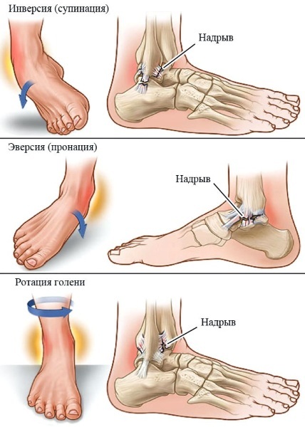
- swelling of the ankle joint, which was formed after mechanical damage to the lower limb (dislocation, fall from a great height, impact on hard-surfaced objects);
- stretching of the connective tissue of the joint, obtained during running, playing sports, brisk walking, as well as due to an incorrectly placed foot;
- fractures and cracks in the tibia, fibula, or talus that come in contact with ankle joint (in this case, an X-ray is necessary to exclude complications and hidden injuries connective tissue);
- objective suspicions of arthrosis, osteoporosis, arthritis, osteosclerosis and other degenerative diseases of this part of the lower limb;
- a feeling of sharp or aching pain, which is localized inside the ankle joint, and also intensifies after a short load or due to a sharp change in weather conditions;
- stiffness of movements in the morning, when, after waking up from sleep, a person needs 10 to 30 minutes to stretch his leg and restore the normal functioning of the ankle (in most cases, such limb dysfunction is accompanied by pain syndrome);
- a radical change in the gait and shape of the foot, which also manifests itself in premature wear of the shoe, atypical deformation of the sole with the curvature of the shoe to the inner or outer side;
- violation of local blood circulation, when the skin surface and soft tissues of the foot are characterized by increased pallor, cyanosis of blood vessels and more low temperature (edema and inflammation inside the ankle can have a static effect on the vessels, disrupting the circulation of the blood channel);
- suspicion of an infectious lesion of the ankle joint by bacterial microorganisms;
- hereditary predisposition to pathological changes in the structure of the ankle, which are manifested in a certain age (X-ray examination is indicated for prophylactic purposes for the timely detection of inflammatory process);
- accumulation of serous exudate, which fills the ankle cavity, causes further destruction of bone and connective tissue (similar pathology may arise due to a previously suffered injury of the lower limb, the consequences of which were not eliminated with the help of medication therapy);
- preparation of the patient for the removal of the destroyed ankle joint with the further installation of an artificial implant or the rehabilitation period after already undergone surgical intervention.
A referral for X-ray examination of the ankle may be ordered by a traumatologist, surgeon, or orthopedist. It all depends on what kind of disease of the musculoskeletal system is found in a particular patient. The course of therapy is formed according to the results of decoding an X-ray image.
How is it determined
Ankle X-ray is the basic examination method used to diagnose this part of the leg. Determination of the pathological state of the ankle or refutation of the presence of the disease is carried out using X-ray equipment.
Fixation of the bone and connective tissue of the joint is carried out by a radiologist in a special office, isolated from the working area of doctors. In the process of diagnostics of the ankle, analog equipment can be used with image display on an X-ray sensitive film, or digital media.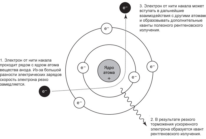
The principle of taking a picture of the ankle joint is that the X-ray beam is directed to the area of the lower leg segment. Passing through the bone and connective tissue of the joint, radiation is scattered unevenly. X-rays pass through less dense tissues, lingering on larger bone structures.
In this regard, a clear image of the bones of the lower limb and all elements of the ankle joint is formed on the X-ray sensitive film. Soft tissues form only a barely noticeable shadow; the human skeleton can be viewed without additional equipment.
Analogue X-ray machines are still used in most public hospitals. These devices record the captured image on a cassette with a sensitive film.
To view such a picture, the use of a special stand with illumination is required. Modern X-ray machines are equipped with an electronic matrix and are connected to a computer monitor.
After a picture is taken with the help of a stream of X-ray radiation, an image of the ankle joint is displayed on the screen of an electronic device. The radiologist gets the opportunity to study the condition of the joint in real time.
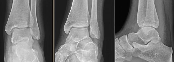
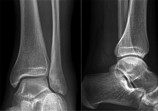
Further storage of the image is carried out in digital format. If necessary, the image of the ankle is printed on paper. To obtain objective information about the state of health of all elements of the joint, an X-ray examination is shown in 2 projections.
First, the patient is asked to rotate the foot so that the front part of the limb and ankle joint can be fixed. In this case, the condition of the talus and tibia bones, the frontal segment of the ankle is assessed.
This type of X-ray examination is especially important if there is a suspicion of combined injury of the ankle joint with fracture of the tibia or elements of the anterior part feet.
A lateral ankle X-ray is performed while lying down. This type of research is carried out in order to comprehensively study the condition of the limb, to fix not only the ankle, but also the calcaneus, fibula, metatarsal bones. These elements of the musculoskeletal system can also be damaged during an ankle injury.
Depending on the type of disease, the attending physician may decide to prescribe an ankle X-ray using a load. In this case, forceful flexion of the foot is performed, taking a picture in a standing position exclusively on the sore limb.
When using these types of x-rays, it is allowed to use local anesthetics. For example, if a person suffers from acute inflammation of the ankle.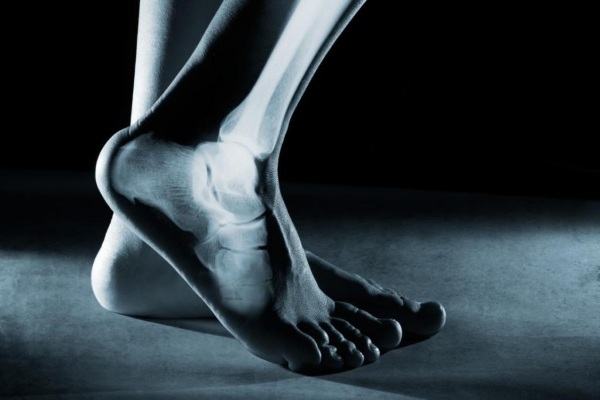
If in the process of X-ray examination of the joint it is established that this diagnostic method does not provide comprehensive information about the condition ankle, the attending physician prescribes an additional examination using the following equipment:
- MRI of the lower limb on which the affected joint is located;
- Ultrasound;
- computer radiography.
All of the above research methods are non-invasive, do not cause pain, last no more than 5-10 minutes, but allow obtain a three-dimensional image of the ankle, bones and connective tissues located in the immediate vicinity joint.
Preparation and analysis
Ankle X-ray does not require complex preparatory measures.
It is enough to observe the following recommendations:
- 48 hours before the appointed date of the diagnostic study, provide the diseased limb with complete rest, excluding any physical activity, the presence of which may affect the results of the examination;
- 24 hours before visiting a doctor, do not take non-steroidal anti-inflammatory drugs, as well as other drugs that have symptomatic therapy (if the patient is undergoing self-treatment with tablets, ointments or gels, then this fact must be notified doctor);
- protect the sore joint from the effects of cold air by putting on a warm sock on its surface or wrapping it with a woolen cloth (the exception is those cases when the pathological condition of the ankle is associated with mechanical damage to its tissues as a result of injury);
- before going to the doctor, you should not wear too tight shoes on the sore leg, which squeeze the joint, disrupting the local blood circulation.
It is necessary to go to the examination together with a relative or close friend who will help to reach to the X-ray room, as well as provide support in the event of acute pain in the ankle joint.
In the complete absence of the possibility of independent movement, it is recommended to call an ambulance. Fulfillment of the above training rules is relevant in the context of a routine examination, when a person is sick arthritis or began to feel attacks of periodic pain in the joint, felt a decrease in its functional activity.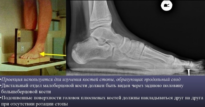
Ankle X-ray is as follows:
- The patient enters the room for the X-ray examination.
- Then he puts his foot in a strictly marked place opposite the diagnostic equipment.
- The doctor turns on the X-ray machine and directs its rays to the ankle joint. The image of the front part of the limb is captured.
- After that, the patient takes a lying position, presses the healthy leg to the chest, and the sore ankle remains on the surface of the couch.
- The doctor takes a lateral x-ray of the joint.
Upon completion of the diagnostic procedure, the image of the diseased limb is displayed on an X-ray sensitive film or recorded on an electronic carrier. It all depends on the type of medical equipment that was used during the examination. The average cost of diagnostics is 2200 rubles.
Decoding the results
The process of decoding an X-ray image should only be carried out by a radiologist, surgeon, traumatologist or orthopedist. The interpretation of the results obtained is carried out by comparing the obtained image of the ankle joint with the norm indicators.
The doctor performs a comparative characteristic of the lower limb of a healthy person with an X-ray image of the patient. Based on the results of the examination, he makes a final diagnosis with confirmation of the joint disease, or refutation of suspicions of pathology of the musculoskeletal system.
The table below lists the main criteria for assessing the health of the joint, which can be used to self-decipher an X-ray image.
| Comparative criteria | Interpretation of results |
| Hyperostosis or atrophy | When examining the image of the ankle, increased attention is paid to the location of the bones, their physiological size and shape. The presence of additional growths or a change in the position of individual elements of the joint is a deviation from the norm and can indicate a chronic or acute inflammatory process, the consequences of which are bone atrophy. Especially if, in addition to this, the patient constantly or periodically feels aching pain in the joint. |
| Structural changes in bone and connective tissue | Comparative characteristics of structural changes in the bone and connective tissue of the joint is aimed at detecting diseases such as osteoporosis and osteosclerosis. In the first case, the picture clearly shows that the bones of the ankle acquire a porous structure, and the process of their demineralization takes place. As a result, the joint becomes more fragile, painful, prone to frequent injury and inflammation. With osteosclerosis, a completely opposite process is observed. The density of the bone and connective tissue of the joint increases. The risk of the formation of extraneous growths that disrupt the functionality of the limb increases. |
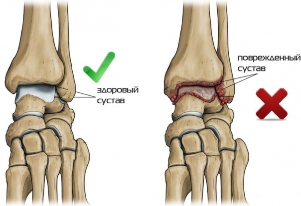
| Joint gap | In a person with a healthy ankle, the joint space has the same lumen width, which can contract slightly under the influence of compression pressure. For example, if an X-ray is taken of a joint under stress. Uneven narrowing of the joint space indicates the presence of the first signs of developing arthrosis or arthritis. At the later stages of this disease, connective tissue is formed in the cavity of this lumen, the formation of which is due to chronic inflammation. Ultimately, the joint no longer functions, and the person is deprived of the ability to move independently with flexion of the foot. |
| Bone surface | Mechanical injuries of the ankle, dislocations, cracks and fractures can lead to the development of degenerative processes in the composition of bone tissue. The presence of signs of ossification or detachment of individual elements of the joint is a pathology that requires complex treatment. |
| Extraneous neoplasms | The structure of the ankle joint can be affected by neoplasms of benign or oncological origin. The appearance of extraneous elevations, additional elements of a round or oval shape, which are part of the bone, indicates the possible presence of a tumor. The type of neoplasm is determined by conducting additional studies in the form of a biopsy of bone tissue samples. |
In the process of comparing the radiographic image of the joint with the image of the ankle of a healthy person, all of the above criteria must be taken into account. At the slightest suspicion of a degenerative, inflammatory or neoplastic process, you should seek medical help.
When to see a doctor
A visit to a doctor should take place in the first 24 hours after a person has felt the following symptoms indicating a pathological condition of the ankle joint:
- sharp or aching pain that worsens at night, after minor physical exertion or a change in weather conditions;
- swelling of the ankle of unknown etiology;
- contusion or dislocation of the foot resulting from a fall, jump from a great height or strong impact;
- a change in the color tone of the skin in the area of the joint (redness or cyanosis of the epithelial tissues indicates inflammation of the bones);
- increase in local body temperature;
- the formation of fistulous canals with the appearance of serous exudate, which is secreted from the skin in the area of the ankle (this symptom is accompanied by attacks of acute pain);
- loss of joint mobility, attempts to restore which lead to pain.
If you have the above symptoms, you should consult a traumatologist or surgeon. Unauthorized treatment with the use of medication therapy, compresses and physiotherapy massage will not bring a positive effect, and can also cause a deterioration in well-being.
Possible complications
People with ankle disease who do not see a doctor and do not deal with the treatment of pathology may face the following complications of the disease:
- purulent inflammation of the bone or connective tissue of the joint, the elimination of which requires surgery and the use of potent antibiotics;
- destruction of the structural elements of the ankle, which are responsible for the process of flexion of the limb;
- the formation of cartilage tissue in the cavity of the joint space, which also deprives the leg of full mobility;
- severe form of arthritis or arthrosis of the joint, accompanied by attacks of excruciating pain, as well as tissue deformation;
- progressive osteomyelitis with bacterial damage to the soft tissues, cartilage and bones of the joint (this complication is the most dangerous and can lead to amputation of the limb).
X-ray examination of the ankle joint is an informative diagnostic method that is most often used in the process of detecting diseases of this part of the leg. The survey is carried out in public or private health care institutions. Ankle X-rays are prescribed by a traumatologist, surgeon, or orthopedist.
This type of diagnosis allows you to timely determine such diseases as arthritis, osteoporosis, arthrosis, osteosclerosis, inflammation of connective and bone tissue caused by bruises, dislocations and other injuries limbs. Decoding of an X-ray image is carried out by conducting a comparative analysis with an image, which is the standard of the norm.
Ankle X-ray Video
How an x-ray is done:


