
Cranial nerves They transmit some of the information from the senses to the brain and connect it with muscles and internal organs, which enables a person to perform various actions of daily life. A table with the characteristics of all 12 pairs is presented in the article.
Record content:
- 1 What are the cranial or cranial nerves?
-
2 Classification
- 2.1 By the place of exit
- 2.2 By function
-
3 12 pairs of cranial nerves
- 3.1 Olfactory nerve (I pair of cranial nerves)
- 3.2 Optic nerve (II pair of cranial nerves)
- 3.3 Oculomotor nerve (III pair of cranial nerves)
- 3.4 Block nerve (IV pair of cranial nerves)
- 3.5 Trigeminal nerve (V pair of cranial nerves)
- 3.6 Abducens nerve (VI pair of cranial nerves)
- 3.7 Facial nerve (VII pair of cranial nerves)
- 3.8 The vestibular cochlear nerve (VIII pair of cranial nerves)
- 3.9 Glossopharyngeal nerve (IX pair of cranial nerves)
- 3.10 Vagus nerve (X pair of cranial nerves)
- 3.11 Accessory nerve (XI pair of cranial nerves)
- 3.12 Hyoid nerve (XII pair of cranial nerves
- 4 What can damage to each pair of nerves lead to?
- 5 Cranial Nerves Videos
What are the cranial or cranial nerves?
A nerve or neuron is the basic structural and functional unit of the nervous system that facilitates communication and signal transmission throughout the body. The brain is one of the main parts of the central nervous system, which is located inside the skull. Hence, the nerves emanating from the brain are cranial.
They are mainly associated with the head and neck and are involved in the transmission of sensory and motor information from the brain to the head, neck, face. This information triggers bodily reactions, helps "feel" certain sensations, and tells the body what to do.
Cranial nerves, 12 pairs (table, functions, characteristics are presented below) belong to the somatic nervous system.
Classification
The cranial nerves supply sensory, motor and autonomic innervation to the structures of the head and neck. They are classified according to their exit from the skull and functionally.
By the place of exit
All cranial nerves exit the brain, bypassing the spinal cord. They are part of the peripheral NS, as they are directed to the cranial and cervical structures. Sensory nerves are formed from young neuronal cells, and motor nerves are formed from cells of the motor nuclei.
| Department of the brain | Associated cranial neuron |
| Brain | I |
| Thalamus | II |
| Midbrain | III IV V VI Vii VIII (partially) |
| Medulla | VIII (partially) IX X XI XII |
Once released, the nerves travel from their source to various parts of the body: the head, face, mouth, and some along the periphery of the body.
By function
The cranial nerves (12 pairs of the table characterize for reference) are classified as sensory, motor, or a combination of both.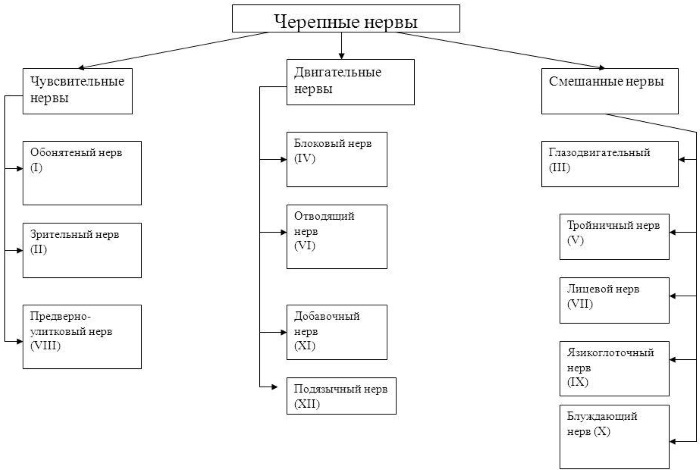
This means that the axons (neurites that carry nerve impulses) in these nerves originate from sensory ganglia (a ganglion of axons) outside the skull or motor nuclei in the brain stem. Axons with sensory functions form synaptic endings, with which they subsequently enter the stem nuclei.
| Sensory | Olfactory, visual, vestibular-cochlear pair | The first 2 pairs, the olfactory and optic nerves, develop as outgrowths in the anterior cerebral bladder. These nerves do not have peripheral sensory nodes. |
| Motor | Oculomotor pair, trochlear nerve, abducens, accessory and hypoglossal nerves | Motor axons innervate the skeletal muscles of the head or neck. |
| Mixed (nerves of the branchial arches) | Trigeminal, facial, glossopharyngeal, vagus | The sensitive parts of the nerves of the branchial arches have nerve nodes (ganglia) in which the organs of peripheral sensory neurons are located. |
The cranial nerve nuclei are functionally organized into separate nuclei in the brainstem. Typically, the nuclei closer to the back tend to be sensory, while the nuclei located in the front tend to have motor functions.
12 pairs of cranial nerves
The cranial nerves are presented in the form of a table. All 12 pairs are numbered with Roman numerals from I to XII according to their location on the brain stem, from the top to the bottom, then from the medial to the lateral and in the order of their exit from the skull.
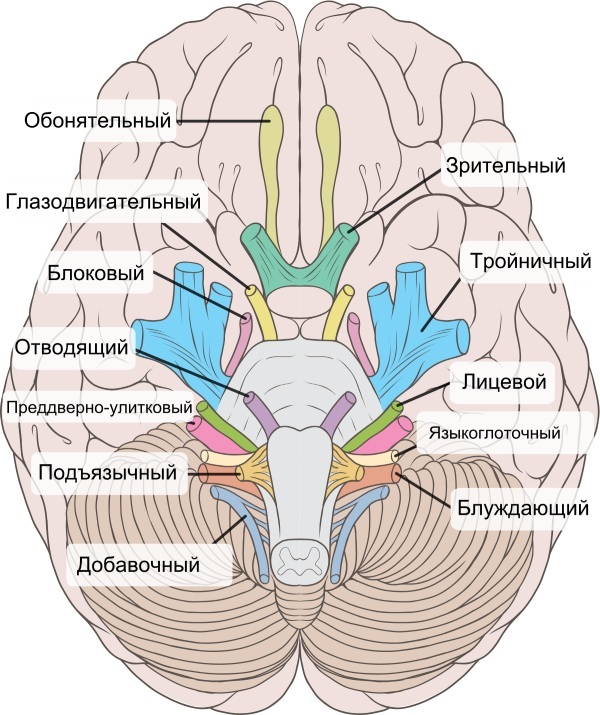
They come in pairs, one on each side of the brain. Each pair serves a specific purpose and has either a motor function, a sensory function, or both.
Olfactory nerve (I pair of cranial nerves)
This is a sensory neuron responsible for the sense of smell, the receptors of which detect smells, transmit information to the brain, where it is responsible for recognizing them.
The olfactory receptors are located within the nasal epithelium. Their axons gather in small bundles of true olfactory nerves that penetrate the small openings of the ethmoid plate of the ethmoid bone and enter the cranial cavity. Once there, the fibers penetrate the olfactory bulb, which lies in the olfactory groove within the anterior cranial fossa.
The olfactory bulb is an ovoid structure that contains specialized neurons called mitral cells. The fibers of the olfactory nerve connect with mitral cells to form clusters known as synaptic glomeruli. Then, from the glomeruli, second-order nerves pass posteriorly into the olfactory tract.
The sensory function of the olfactory neuron is achieved through the olfactory mucosa. This layer of the shell not only senses the smell, but also determines the more complex aspects of the taste.
Optic nerve (II pair of cranial nerves)
Pair II is the sensory nerves that lead to the optic junction and carry information to the occipital lobe.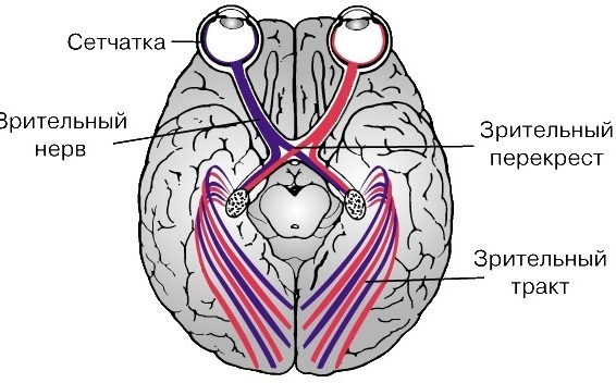
The visual pair is located at the back of the eyes. The work consists in transmitting visual information from the retina to the visual centers of the brain using electrical impulses.
The structure is composed of ganglion or nerve cells. Thanks to this nerve, sensory nerve impulses are transmitted from more than 1 million retinal ganglion cells to the visual centers of the brain. The vast majority of optic nerve fibers transmit information about central vision.
The nerve begins at the disc (structure, 1.5 mm in diameter) at the back of the eye. The disc is formed by the convergence of the exit fibers of the ganglion cells (axons) as they exit the eye. After exiting it, it passes through the orbit, the bony canal of the optic nerve, and exits intracranially on the underside of the front of the brain.
At this point, the optic nerve in each eye connects and forms an X-shaped structure called the optic chiasm. Here, about half of the nerve fibers from each eye continue on one side of the brain, and the remaining nerve fibers intersect to join fibers from the opposite eye on the other side brain.
This arrangement is necessary to obtain binocular vision. Behind the intersection, fibers travel along the visual pathways to various parts of the brain. Certain nerve fibers leave the optic tract and enter the brain stem to provide information that ultimately determines the size of the pupil.
Oculomotor nerve (III pair of cranial nerves)
The oculomotor lives up to its name, helping to move the eye and focus on objects through pupil control. It is also responsible for keeping the eyelids open by innervating the muscles of the upper eyelids.
The pair develops from the large-cell nucleus, leaves the cranial cavity through the superior orbital fissure. The nerve nucleus does not consist of a continuous row of cells, but is divided into several smaller nuclei, which are divided into 2 groups: anterior and posterior.
Each of them controls a specific muscle. In the posterior group, there are 6 of them, 5 of which are symmetrical on both sides of the midline, and the 6th is located in the center and is common to the nerves on both sides.
There are sympathetic fibers that run along the superior branch of the oculomotor nerve. They innervate the superior tarsus muscle, which keeps the eyelid lifted.
Block nerve (IV pair of cranial nerves)
The trochlear is one of the smallest motor nerves that controls the superior oblique muscles of the eye.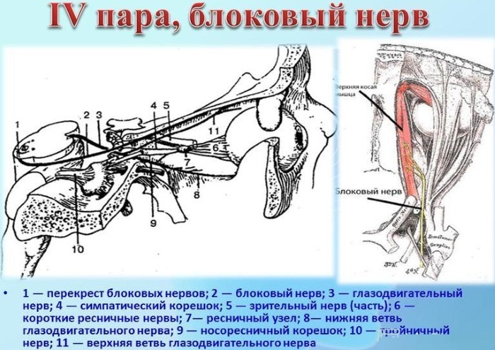
It has the smallest number of axons. It is a nerve with motor and somatic functions associated with the superior oblique muscle, allowing the eyeball to rotate. The nuclei of the block nerve are also associated with the midbrain,
The nerve arises from the block of the nucleus of the brain that emerges from the posterior part of the midbrain (this is the only cranial nerve that emerges from the posterior part of the midbrain). Passes anteriorly and inferiorly in the subarachnoid space, then moves along the lateral wall of the cavernous sinus before entering the orbit of the eye through the superior palpebral fissure.
This nerve is the thinnest of all cranial neurons and has the longest intracranial pathway.
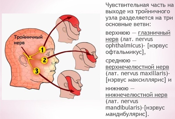
Trigeminal nerve (V pair of cranial nerves)
Cranial nerves (12 pairs of the table characterizes in general terms), which depart from the lower basal surfaces of the brain include nerves that perform sensory and motor functions at the same time. These include the trigeminal nerve. It splits into 3 main branches after the trigeminal ganglion.
The eye branch transmits sensory information from:
- heads;
- forehead;
- the upper part of the sinuses;
- upper eyelid and associated mucous membranes;
- the cornea of the eye;
- bridge of the nose.
The maxillary region transmits sensory information from:
- lower eyelid and associated mucous membranes:
- the middle part of the sinuses;
- nasal cavity;
- cheeks;
- upper lip;
- some of the teeth of the upper jaw and associated mucous membranes;
- palate.
The mandibular region is the only part of the trigeminal nerve that performs both sensory and motor functions.
These include:
- the outer part of the ear;
- the lower part of the mouth and associated mucous membranes;
- the front and middle of the tongue;
- lower jaw teeth and associated mucous membranes;
- underlip;
- the chin.
The trigeminal nerve also stimulates movement of the muscles in the jaw and some muscles in the inner ear. It originates from 3 sensory nuclei and one motor nucleus extending from the midbrain to the pons. At the level of the medullary bridge, the sensory nuclei merge to form the sensory root. In the middle cranial fossa, the sensory root expands to the trigeminal ganglion.
The peripheral aspect of the ganglion is subdivided into 3 sections:
- ocular;
- maxillary;
- mandibular.
The motor root runs below the sensory root along the bottom of the Meckel Cave. Its fibers extend only to the mandibular region.
The maxillary nerve leaves the skull through the superior orbital fissure and round foramen. The mandibular exits through the foramen ovale and enters the temporal fossa.
Abducens nerve (VI pair of cranial nerves)
It is a motor nerve that controls eye movement. It performs a somatic motor function - it provides innervation to the lateral rectus muscle.
The abducens nerve leaves the nucleus of the same name at the junction of the bridge of the trunk and the medulla oblongata. It then enters the subarachnoid space and penetrates the dura mater to enter an area known as the Dorello canal. At the end of the petrous temporal bone, the abducent nerve exits the canal and enters the cavernous sinus (venous sinus of the dura mater).
It passes through it and enters the bony orbit through the superior orbital fissure. Within the bony orbit, the neuron innervates the lateral rectus muscle of the eye, which is responsible for opening and closing the eye.
Facial nerve (VII pair of cranial nerves)
This pair performs both sensory and motor functions. The facial nerve is associated with derivatives of the second pharyngeal arch, innervates the facial muscles, the posterior part of the digastric abdomen, the stylohyoid and stapes muscles.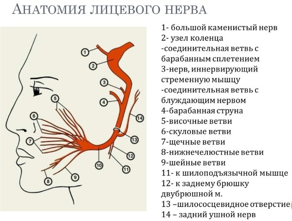
Sensory perception:
- a small area around the auricle;
- provides a special taste sensation to the front 2/3 of the tongue.
Parasympathetic perception:
- submandibular and sublingual salivary glands;
- mucous glands of the nose, palate and pharynx;
- lacrimal glands.
The course of the facial nerve is very difficult. There are many branches that transmit a combination of sensory, motor, and parasympathetic fibers.
Anatomically, the stroke can be divided into 2 parts:
- Intracranial passes through the cranial cavity and the skull itself.
- The extracranial goes outside the skull, through the face and neck.
The intracranial nerve is formed in the region of the brainstem. It starts as 2 roots: large motor and small sensory. Both pass through the internal auditory canal, leave it, and enter the facial canal. Inside the canal, first 2 roots merge, forming the facial nerve, which then forms the geniculate node (a collection of nerve cell bodies).
Here the nerve gives rise to:
- large stony nerve;
- stirrup;
- drum chord.
Then the facial nerve leaves the skull upward, passes just in front of the outer ear, where it provides motor innervation to some muscles.
Inside the parotid gland, it divides into 5 branches:
- temporal;
- zygomatic;
- cheek;
- marginal mandibular;
- cervical branch.
These branches are responsible for the innervation of the facial muscles, some muscles of the head and neck.
The vestibular cochlear nerve (VIII pair of cranial nerves)
The sensory nerve, which is responsible for hearing and balance. It consists of 2 parts: vestibular and cochlear, which originate from different nuclei. Both have a sensory function.
- The vestibular component arises from the complex of vestibular nuclei in the pons and medulla.
- The cochlear component arises from the ventral and dorsal nuclei of the cochlea, located in the inferior cerebellar peduncle.
Both types of fibers are connected in the bridge, forming the vestibulocochlear nerve. It exits the brain at the cerebellopontine angle, and from the skull through the internal auditory canal of the temporal bone.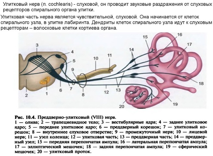
In its distal part, the nerve is divided, forming the vestibular and cochlear processes. The first innervates the vestibular system, which is responsible for determining the balance. The second goes to the cochlea of the inner ear, forming spiral ganglia, which are responsible for hearing.
Glossopharyngeal nerve (IX pair of cranial nerves)
It is both a motor and sensory nerve, which is responsible for sending sensory information to the sinuses, throat, ear, and tongue. It is also responsible for the movement of the muscles in the back of the throat. It also innervates the chemoreceptors in the carotid artery and the baroreceptors of the blood pressure sensor in the carotid sinus. Its visceral motor function is salivation through the parotid gland.
The nerve starts in the medulla oblongata. Moving in the posterior cranial fossa, it leaves the skull through the jugular foramen, at the exit there is a tympanic nerve that penetrates the temporal bone and enters the middle ear cavity.
Here it forms the tympanic plexus - a network of nerves that provides sensory innervation:
- middle ear;
- the inner surface of the tympanic membrane;
- eustachian tube.
At the level of the stylopharyngeal muscle, a nerve of the carotid sinus is formed, which descends along the neck, innervating the carotid sinus and the body of the carotid artery.
The glossopharyngeal nerve ends with a division into several sensitive branches:
- pharyngeal - combines with the fibers of the vagus nerve, forming the pharyngeal plexus, innervates the mucous membrane of the oropharynx;
- lingual - provides a general and gustatory sensation of the posterior third of the tongue;
- tonsillar - forms a network of nerves known as the amygdala, which innervates the tonsils.
Vagus nerve (X pair of cranial nerves)
It is a motor and sensory nerve that:
- responsible for the taste of the tongue;
- controls the throat muscles, organs of the ear canal;
- sends information to the heart and intestines;
- stimulates muscle movement in the digestive tract.
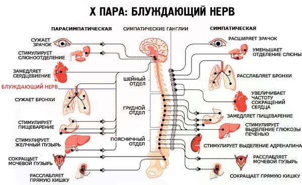
The vagus nerve has the longest path of all cranial neurons, extending from the head to the abdomen. Its name comes from the Latin "vagary", which means "wandering."
It originates from the medulla oblongata, leaves the skull through the jugular foramen, through the glossopharyngeal and accessory nerves. An ear branch appears inside the skull. This provides sensitivity to the back of the external auditory canal and external ear. In the neck it passes into the carotid sheath, below the internal jugular vein and the common carotid artery.
Several branches appear on the neck:
- Branches of the pharynx - provide motor innervation of most of the muscles of the pharynx and soft palate.
- Superior laryngeal nerve - is divided into internal and external branches. The external laryngeal innervates the cricoid muscle of the larynx. The inner larynx provides sensory innervation to the larynx and the upper larynx.
- Recurrent laryngeal nerve (right side only) - hooks under the right subclavian artery, then rise to the larynx. It innervates most of the internal muscles of the larynx.
At the base of the neck, the right and left nerves travel in different ways into the chest, forming the posterior and anterior trunk of the vagus nerve. These branches contribute to the formation of the esophageal plexus, which innervates the smooth muscle of the esophagus.
Two other branches arise in the chest:
- Left recurrent laryngeal nerve - to innervate most of the internal muscles of the larynx.
- Heart branches - they regulate the heart rate, provide the internal sensitivity of the organ. In the abdominal cavity, the trunks of the vagus nerve end with division into branches supplying the esophagus, stomach, small intestine and large intestine (up to the bend of the spleen). Its function is to stimulate smooth muscle contraction and glandular secretion in these organs. For example, in the stomach, it increases the rate of its emptying, stimulates the production of acid.
Accessory nerve (XI pair of cranial nerves)
It is a motor nerve that controls the muscles in the neck. Unlike other cranial nerves, it consists of 2 parts: the cranial, which arises from the medulla, and the dorsal, which arises from the cervical region. The dorsal portion rises through the opening and connects to the cranial portion to form an accessory nerve. It has only motor fibers and is divided into the dorsal and cranial regions.
It innervates:
- Sternocleidomastoid muscle. It runs from the mastoid process of the temporal bone to the head of the sternum and clavicle. Helps to carry out lateral flexion and rotation of the neck in unilateral action, and its extension in the atlanto-occipital joints in bilateral action.
- Trapezius muscle. Consists of top, middle and bottom fibers. The upper fibers of the trapezium raise the scapula and rotate it when the arm is abducted. The middle fibers pull the scapula in, and the lower ones pull it down.
Hyoid nerve (XII pair of cranial nerves
The cranial nerves, 12 pairs (the table for convenience is presented on the website) of which are responsible for the work of the whole organism, also include the hypoglossal nerves. They belong to the motor structures, control the movement of the tongue. They supply the motor fibers to all muscles of the tongue, except for one - the palatine-lingual muscle, which depends on the vagus nerve.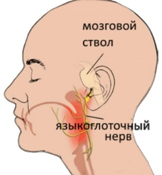
The neuron arises from the hypoglossal nucleus in the brainstem. It then passes through the posterior fossa and exits the skull through the hyoid canal. Here it takes a branch of the cervical plexus, along which fibers from the roots of the spinal nerves pass. It then passes below the angle of the mandible, crosses the internal and external carotid arteries, and travels forward into the tongue.
The nerve provides motor innervation to most of the muscles in the tongue. Another branch, containing fibers, descends to supply the nerve loop, which is part of the cervical plexus.
What can damage to each pair of nerves lead to?
Damage to the cranial nerves, their tracts or nuclei leads to dysfunctions:
- If the facial nerve is damaged, one side of the face will not be able to express emotions.
- The optic nerve can be damaged by skull fractures. In this case, irreversible blindness of the affected eye may develop.
- Any damage or trauma to the vestibular cochlear structure can result in hearing loss or imbalance.
- Injury to the hypoglossal nerve can lead to complete paralysis of the tongue, as a result of which a person cannot eat or speak normally. There are many reasons that can damage the hypoglossal nerve, such as an infection or nerve damage that leaves the tongue paralyzed.
- Any dysfunction of the accessory nerve can cause the shoulders and neck to not work properly.
- Damage to the abducens nerve can lead to blurred vision or double vision.
- If the vagus nerve is damaged, the resting heart rate can be around 100 beats per minute.
Cranial nerves, 12 pairs (the table is presented in the article for review) which are located inside the skull, when damaged, require focusing on protecting the brain. Some of the treatments for their diseases include surgery. Tumors are treated with radio waves.
The cranial nerves carry information from the brain to parts of the human body, especially the head and neck, and vice versa. Many of the 12 pairs are connected by connecting branches. The structures and connections of the human body are presented in anatomical tables and atlases, which help to better understand the work of the body.
Author: Belyaeva Anna
Cranial Nerves Videos
Cranial nerves in 1 minute:


