
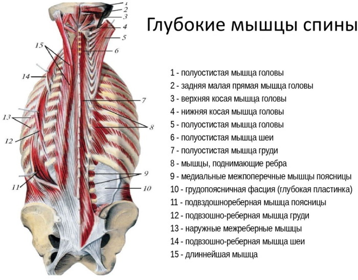
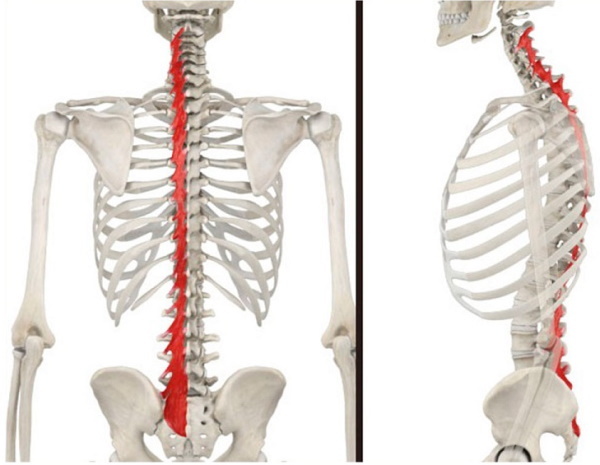
On the back of the body there are 3 layers of muscles that are connected to the spine and run from the pelvis to the head. These deep back muscles or transverse spinous, which are largely responsible for maintaining posture and spinal mobility.
Record content:
- 1 Characteristic
- 2 Functions and properties
-
3 Structure
-
3.1 Semispinal muscle
- 3.1.1 Breasts
- 3.1.2 Neck
- 3.1.3 Head
- 3.2 Multipart muscles
- 3.3 Rotator muscles
-
3.1 Semispinal muscle
- 4 Innervation
- 5 Possible diseases and pathologies
- 6 Massage
- 7 Back Muscles Videos
Characteristic
On the back, thick muscle threads run from the pelvis to the head, located on both sides of the spine. They are especially important for stability. Distinguish between superficial and deeper (autochthonous) muscles of the back. The deep, erect spine attaches directly to the spine and keeps it upright.
Muscles form a complex system of tension: how complex this system is, show the various points of muscle attachment. Each spinous process of the vertebral body is connected by muscle groups with several transverse processes, and vice versa, each transverse process is associated with different spinous processes of the vertebral body.
Muscles lie on top of each other in several layers. First short and then long. Thus, they form a strong muscle network that stabilizes the spine, but allows for various movements. The longitudinal muscles can tilt the body back or sideways, the oblique muscles can rotate it.
The compact muscle network surrounding the spine is designed to move and support the spinal column. The muscles closest to the spine are the deep ones, which stabilize it.
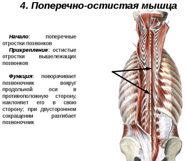
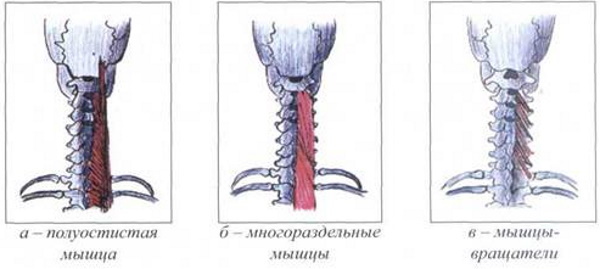
The transverse spinous muscle of the back is 3 groups of deep muscles:
- Semispinalis.
- Multifidus.
- Rotatores.
They form the transversospinalis, and, in fact, unite the spine in many different ways, connecting the transverse processes with the spinous in various ways. Together, they affect stability, movement, and proprioception.
 Although they are separate muscles, in fact, they form a chain that requires each link to be active and participate in working together. Poor posture, especially in the form of a backward bend, makes it very difficult to get all of these muscles to work together as a team.
Functions and properties
The load-bearing element of the human skeleton does not pass exactly through the middle of the body. This means that the center of gravity of the body mass does not coincide with the spine. In order for a person to be able to straighten up and not immediately fall under the weight of the chest, abdomen and intestines, he needs a complex network of muscles that would balance the forces of gravity.
In addition, deep muscles are involved in all movements of the spine, for example, when bending back or to the sides, and turning to the right or left. The internal spinal muscles receive a signal to move from the branches of the spinal nerves to the spinal cord.
| Transverse spinous muscles | Functions |
| Semispinalis | Bilateral contraction: extension of the head, thoracic spine and cervical. Unilateral: lateral flexion and rotation of the head, thoracic, cervical spine. |
| Rotatores | Bilateral contraction: extension of the thoracic spine. Unilateral: rotation of the thoracic spine. |
| Multifidus | Bilateral contraction: extension of the spine. Unilateral with: lateral flexion and rotation of the spine. |
In addition to deep muscles, which are attached directly to the spine, there are superficial muscles in the back. They connect the spine to the chest, head, arms and legs. They can be trained directly, as opposed to deep muscles, which can only be trained indirectly.
Structure
The transverse spinous muscle of the back is the generic name for the dorsal muscles of the transversospinales. It is represented by muscle bundles that have different lengths and are thrown over a different number of vertebrae.
In this case, separate, layer-by-layer muscles are formed:
- peninsular;
- multipartite;
- rotational.
All 3 types are part of the transversospinalis group (transverso - transverse, spina - spine). Tiny individual muscles connect the transverse process of one vertebra to the spinous process of another, located above.
This muscle network, located at the back of the torso, rotates the spinal column, pulling the spinous processes down towards the associated transverse processes.
All muscles of this group are located very close to individual intervertebral joints. This proximity allows them to control the joints, stabilize them, since the larger superficial muscles act on the trunk.
The deep muscles of the spine, although not all connected to each other, tend to work in a chain. All muscles follow different attachment patterns: some cross one vertebra, and others 2 or more, although none crosses more than 6. Poor posture often leads to chain breakage and misalignment in this area.
Semispinal muscle
The Semispinalis back muscle is the most superficial of the transversospinalis muscle group. Its fibers connect each transverse process to the spinous process five or six vertebrae above. The direction of the fibers is the most vertical in the group, which gives the best opportunities for spinal extension.
The semispinal muscles are located on the sides of the spine and are divided into 3 muscles: chest, neck and head. They attach to the transverse processes (bony protrusions from the arch of each vertebra) of the lower vertebrae and travel upward through several vertebrae to reattach.
These are the most superficial of the group of transverse spinous muscles. The places of their attachment are the thoracic, cervical vertebrae and the occipital bone at the base of the skull.
Breasts
The semispinalis muscle of the chest (Semispinalis thoracis) has thin bundles with long tendons extending from both ends. They arise from the transverse processes of 6-10 thoracic vertebrae (T6 - T10). Tendons are inserted into the spinous processes from the 6th cervical to the 4th thoracic vertebrae (C6 - T4).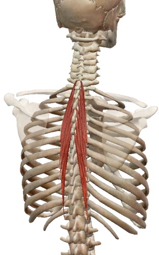
When contracted on both sides, the pectoralis muscle expands the thoracic spine. When it contracts from one side, it causes lateral flexion of the spine in the same direction, but rotation in the opposite direction.
The semi-pectoral muscle is innervated by the medial branches of the posterior branches of the spinal nerves, and the dorsal branches of the intercostal arteries provide blood supply.
Neck
Semispinalis cervicis, the semi-spinal muscle of the neck, arises from the transverse processes from 1 to 6 of the thoracic vertebrae (T1 - T6). Muscle fibers are inserted into the spinous processes of 2-5 cervical vertebrae (C2 - C5).
By contracting on one side, it helps to rotate the neck in the opposite direction of activation and provides lateral flexion in the same direction. By contracting on both sides, this muscle expands the spine.
The semi-spinal cervical muscle is innervated by the medial branches of the posterior branches of the spinal nerves.
Head
The semispinalis muscle of the head (Semispinalis capitis) is a long, thin muscle located deep in the back of the neck. Muscle fibers are formed from the transverse processes of the 4-7 cervical vertebrae (C4 - C7) and 1-6 thoracic vertebrae (T1-T6). Semispinalis capitis is inserted into the spinous processes of 2-5 cervical vertebrae (C2 - C5).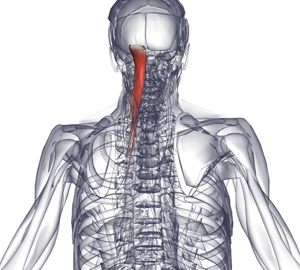
By contracting on both sides, the muscle extends the head and neck, which is a two-way motion. Squeezing on one side, turns the head and neck in the opposite direction and flexes the head and neck laterally in the same direction of activation.
During bilateral contraction, this muscle is considered the main motor for the dynamic maintenance of cervical lordosis. It is also considered to be one of the main factors in maintaining head-to-neck balance.
The muscle is innervated by the descending branches of the greater occipital nerve (C2) and the third cervical nerve (C3). Blood supply is provided by the descending branch of the occipital artery and the superior intercostal artery through the dorsal branches of the two superior posterior intercostal arteries.
Multipart muscles
The transverse spinous muscle of the back consists of several different bundles. Among them there are relatively small ones, such as the multifidus muscles or the Multifidus. They are a group of short triangular muscles.
These are the thickest muscles in the transversospinal group; they are shorter than the semi-spinal muscles, but longer than the rotator ones. Multifidus are found on either side of the spine, extending from the cervical to the lumbar region, and are respectively divided into the cervical, thoracic, and lumbar regions.
- Cervical multifidus muscles arise from the superior articular processes C4 - C7. They extend over the median part and are inserted on the lateral side and at the ends of the spinous processes of the C2 - C5 vertebrae.
- The multiple thoracic vertebrae originate from the transverse processes of the thoracic vertebrae.
- Multifidus fibers in the lumbar region arise from the processes of the lumbar vertebrae and the posterior surface of the sacrum.
The multiple muscles, passing through the groove between the spinous and transverse processes of the vertebrae, overlap the plates of the cervical and thoracic vertebrae and the posterior surface of the sacrum.
The muscles are innervated by the medial branches of the posterior branches of the spinal nerves in the corresponding cervical, thoracic and lumbar regions.
The multifidus receives arterial blood supply from a number of arteries along the length of the spinal column:
- Cervical: vertebral, deep cervical and occipital arteries.
- Thoracic region: dorsal branches of the posterior intercostal and subcostal arteries.
- Lumbar region: lumbar and lateral sacral arteries.
 Each multifidus muscle connects 3 to 6 vertebral levels between the transverse and spinous processes of specific cervical, thoracic, and lumbar vertebrae.
Although the multifidus muscles are small, they help several movements of the spine: with bilateral contraction, they expand the spine, while unilateral contraction promotes lateral flexion of the spine in the same direction and rotation of the spine in the opposite direction side.
Rotator muscles
Rotating muscles, rotators, are a set of short muscles located laterally along the spinal column, attached between the transverse and spinous processes of the thoracic vertebrae. As a result, rotators function as stabilizers, extensors, and rotators of the spine.
Rotatores thoracis are small quadrilateral muscles located between the thoracic vertebrae. There are usually 11 pairs present, but some may be missing at both ends of the thoracic spine.
They are attached as follows:
- Long rotators. They originate from the transverse processes of the thoracic vertebrae and are inserted into the bases of the spinous processes and the plates of the thoracic vertebrae located 2 levels higher.
- Short rotators. They have the same attachments as long rotators, but protrude only 1 level above their origin, so they are shorter.
Rotators occupy the third, deepest layer of the internal muscles of the back. These are the deepest and shortest transversospinal muscles. Innervated by the medial branches of the posterior branches of the spinal nerves. Arterial blood is obtained from the dorsal branches of the posterior intercostal and lumbar arteries.
These muscles are the most important subtype affecting the spine. Bilateral contraction leads to extension of the spine, while unilateral contraction causes contralateral rotation of the thoracic spine.
However, the rotator muscles are in positions with very poor mechanical stress, and studies have shown minimal contribution of structures to the movement of the spine. Instead, they function as important stabilizers of the spinal column, acting as extensible ligaments that adjust their length to support adjacent vertebrae.
Innervation
The transverse spinous muscle of the back has the same nerve supply from the dorsal branches of the spinal nerves.
Possible diseases and pathologies
While the superficial muscles of the back play a role in the movement of the spine, the transversospinalis muscle group provides spinal stability, proprioception and posture.
Degenerative changes are associated with pathology:
-
Herniated lumbar disc. Most herniated discs occur in the lower lumbar spine, especially between 4 and 5 lumbar vertebrae, and between the 5th lumbar vertebra and 1 sacral vertebra (L4-5 and L5-S1).
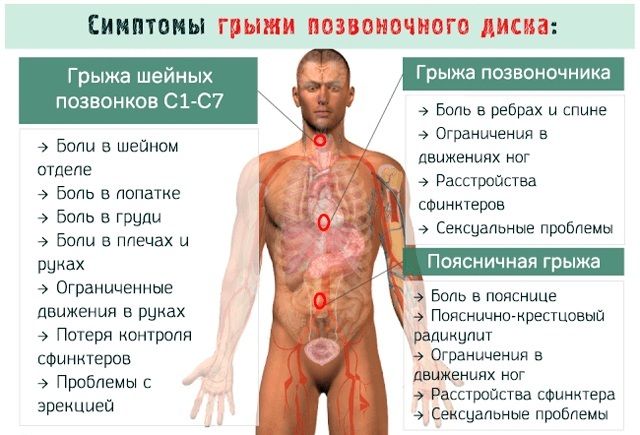
- Osteoarthritis of the facet joints. On either side of each spinous process, there is a facet joint that connects each vertebra together to form the spine. These facet joints are covered with a smooth elastic layer of cartilage that separates the vertebrae and allows their movements to be smooth and painless. Osteoarthritis occurs when a layer of cartilage breaks down and wears away, causing friction between the bones. In more severe cases, painful bone spurs called osteophytes may form at the site of the cartilage. Osteoarthritis is a common cause of back pain. It can develop on its own or in combination with other degenerative conditions of the spine, such as osteochondrosis or spinal stenosis.
Other pathologies include muscle weakness and atrophy. Trigger points in the multifidus muscle reduce the effectiveness of the contraction of the transverse abdominal muscle due to a decrease in reciprocal inhibition (ensuring the coordination of the work of the muscle centers), therefore the disease causes chronic pain in lower back.
Massage
Weakness and imbalance in the muscles of the trunk, neck and throat are considered physical risk factors for back pain. More and more people are spending their day between an office chair, car seat and sofa. This sedentary lifestyle weakens the muscles. Accidental overloading of the spine can easily lead to painful tension.
Weakened core muscles are more likely to cause back pain than herniated discs or wear and tear on bones and joints. Conversely, well-trained back and abdominal muscles are the best protection against back problems. Because strong muscles are elastic and form a natural support corset for bones and joints.
While some types of massage relax and rejuvenate the body, deep tissue massage has a therapeutic effect that can heal the body and restore muscles, tissues and joints. Slow, hard blows to muscle fibers can cause some discomfort because deep tissue massage works on the deep layers of muscle and connective tissue known as fascia.
Benefits of deep tissue massage:
- relieves stress;
- relieves pain;
- improves range of motion;
- lowers heart rate and lowers blood pressure.
To relieve muscle spasms, 4 massage sessions are required over a period of 6 weeks. If muscle spasms do not respond to neuromuscular therapy within 2 sessions, then treatment should be reviewed.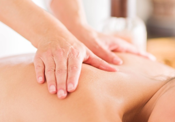
Deep tissue massage technique:
- Massage oil or lotion is applied to the lower back. This makes the skin texture smoother and easier to work with.
- First, the masseur rubs the lower back with his fingertips. It soothes tight muscles and also helps the therapist know which points to work on.
- The rubbing gradually intensifies, it starts from the tailbone or pelvic bone and moves up, in the direction of blood flow. Thus, rubbing brings relief because it increases the flow of fresh blood to the muscles.
- Then, clenching his hand into a fist, the masseur will press only on the focus area. If the knot is strong, the elbows may be used. At this point, you should breathe deeply, because the knots are torn, and the muscles are stretched. However, this should not cause too much pain.
- At the end of the entire process, you should drink a glass of water. If there is a urge to use the toilet soon afterwards, then the session was successful. This is due to the fact that when the nodes in the body rupture, toxins that were previously retained in the muscles are released. Water helps to remove them from the body.
If taking place in a professional spa, you may be asked to take a warm bath after the procedure. This will deepen your relaxation as well as improve hydration and detoxification.
The massage should only be performed by a qualified professional. Lack of knowledge can lead to tissue damage or blood clots.
Poor posture can cause unnecessary pressure and strain on the spine. Over time, this can lead to pain and damage to the back. To prevent this from happening, you should daily strengthen the transverse spinous and many other muscles, do stretching to improve blood circulation and reduce the likelihood of developing back pain.
Back Muscles Videos
Deep Back Muscles Anatomy:


