
Human legs are made up of dozens of muscles. Each one helps to walk, run, jump and bend over. Back of the thigh consists of 3 large muscles, one of which is the biceps femoris. The anatomical structure of these structures is described later in the article.
Record content:
-
1 Features of the anatomy of the biceps femoris
- 1.1 Start
- 1.2 Attachment
- 1.3 Innervation
- 2 Key muscle functions
- 3 Functional muscle tests to assess muscle condition
- 4 Muscle palpation
-
5 Muscle pain causes, possible diseases, symptoms and treatment
- 5.1 Biceps tendon rupture
- 5.2 Tendinitis
- 5.3 Spasm
- 6 Video about the biceps femoris
Features of the anatomy of the biceps femoris
The back of the thigh is covered by 3 large hamstring muscles:
- semitendinosus;
- two-headed;
- semimembranosus muscle.
One end of all the muscles is attached to the lower part of the pelvis and the other to the thighbone in the knee region. The hamstring muscles are responsible for hip extension and knee flexion.
The biceps femoris (human anatomy explains the principles of the dynamism of muscle structures) begins approximately at the point where the buttock area (tailbone) ends and the thigh begins, the large bone of the upper part legs. It then descends to the back of the knee and attaches to the lower leg bones. Biceps are muscles with 2 heads or points of origin.
As the name suggests, the femoral head consists of 2 heads, one of which lies deep within the other. Each head has a different origin and innervation, but the same insertion or location where the muscle ends and attaches. Most of the attachment goes to the fibula, but some parts attach to the tibia, located at the bottom of the leg.
Start
The long head of the muscle lies behind the short one. If you look at the hip from behind, you can see only the long head, and the short one looks out slightly from below. The biceps muscle is the most lateral of the hamstring muscles.

The long head is the strongest muscle in the hamstring group. It arises from the back of the ischium. This tendon is common with the semitendinosus muscle and the sacro-tuberous ligament.
The short head originates from the aspera line (rough ridge of the posterior surface of the thigh) and the supracondylar ridge of the femur. This is about halfway between the thighbone and the knee. Consequently, the short head begins significantly lower in the leg than the long one.
Since the short head does not arise from the ischial tuberosity, it is not considered part of the hamstring. The short head has some anatomical variations in humans, and sometimes is completely absent. The short section extends up the thigh and connects to the gluteus muscles.
The 2 heads form the biceps tendon and are then inserted over the fibular head, the fascia of the thigh, and the proximal tibia.
Attachment
Both parts of the thigh muscle join at the distal thigh to form a 2-head tendon. It attaches to the lateral side of the fibular head and to the lateral condyle of the tibia.
Thus, the 2 heads of the femoral muscle are connected to the lower leg bone on the outside of the leg, and also with the upper and outer surface of the lower leg near where it meets the knee a cup.
For the most part, the biceps muscle passes superficially in the posterolateral part of the thigh, deeply adhering only to the skin, fatty and fascial layer. An exception is its apical part, where it is covered by the gluteus maximus muscle.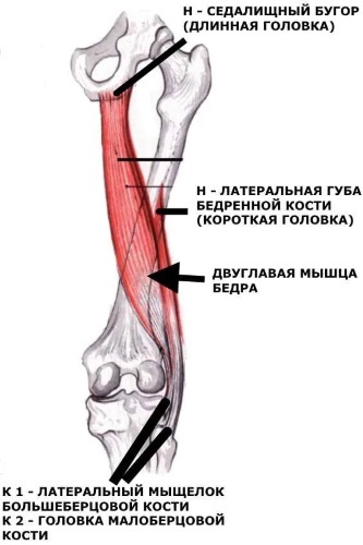
Descending from the pelvis to the back of the thigh, the biceps muscle passes over the semimembranosus, adductor magnus and the lateral head of the gastrocnemius muscle.
Along the way, it is also located superficially in relation to the sciatic nerve, providing it with protection.
The sciatic nerve gives up its terminal branch (common peroneal nerve) near the site of attachment of the biceps femoris.
The nerve briefly travels along the medial border of the biceps muscle, attaching to the tendon. This is important for clinicians when considering trauma or performing surgical procedures in this area.
Innervation
The biceps femoris (anatomy is shown in the pictures for clarity) is supplied with the terminal branches of the sciatic nerve. But 2 heads get innervation from different parts of the nerve.
The large one is innervated from the tibial component of the sciatic nerve, and the small head is innervated from the peroneal part of the sciatic nerve. This unique pattern of innervation can lead to impaired coordination of movements of both heads and vulnerability to sports injury.
The main nutrition of the femoral biceps muscle comes from the branches of the deep femoral artery. Additional comes from the inferior gluteal and superior lateral geniculate artery.
Key muscle functions
A short head and a long one have different effects on muscle action. The first affects only the knee, due to its specific attachment. The long head affects both the thigh and the knees.
The femoral 2-head muscle performs the following actions:
- bends the knee
- turns the leg outward (external rotation);
- straightens the thigh.
The biceps femoris, together with other muscles of the posterior femoral surface, stabilizes the pelvis, especially when the trunk is flexed forward. Hence, it plays an important role in the gait cycle. Anatomically, its main antagonist is the quadriceps femoris muscle.
Functional muscle tests to assess muscle condition
To diagnose the hip joint, various tests are performed, according to the results of which the degree of damage to the hip joints and muscles is determined.
- Faber (Patrick) test means: flexion, abduction and external rotation. The combination of these 3 movements results in a clinical pain provocation test that helps diagnose abnormalities in the hip, lower back and sacroiliac region.
-
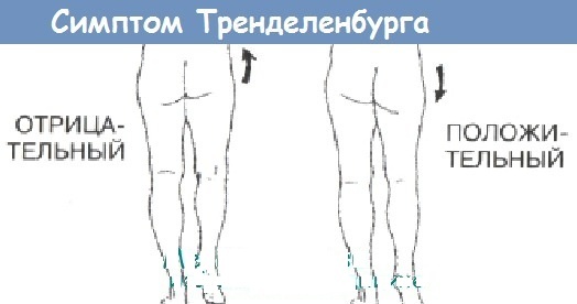
Trendelenburg test Is a quick physical examination that helps the therapist identify any hip dysfunction. A positive test result indicates weakness in the abductor muscles of the thigh: gluteus medius and gluteus minimus. These data can be associated with various pathologies, such as congenital dislocation of the hip, rheumatoid arthritis, osteoarthritis.
- Thomas test used to measure the flexibility of the hip flexors. Impaired range of motion of the hip may be a major cause of other conditions such as pain syndrome in the patella or intercondylar sulcus of the thigh, back pain, osteoarthritis and rheumatoid arthritis.
Muscle palpation
In order to better determine the location of the posterior thigh muscle group, you should first find some points:
- Ischium. It is easily palpable in a sitting position. It is located in the middle of the buttocks, at the level of the gluteal fold.
- Inner knee bend (popliteal fossa). The site of distant attachment of the semitendinosus muscle. The bend of the knee forms the medial part of the popliteal fossa, and the lateral part will be the tendon of the 2-head muscle. With a slow straightening of the lower leg, the soft semimembranous muscle, located deeper than the semitendinosus, is well felt. In this place, the biceps muscle is felt especially well.
Palpation is performed to determine the source of the pain in the hip and assess the pain. In addition to palpation of the painful area, the sacral covering of the spine, ischial tubercles, acetabulum, and abdominal muscles should be checked. Based on the general picture, a diagnosis is made.
Approximate palpation is performed while lying on the stomach or back. In the prone position, the legs should be slightly bent at the knees.
First, the doctor determines the lateral and proximal boundaries of the popliteal fossa, and then, moving the palm along the large muscular abdomens, determines the general consistency of the muscles. At the same time, hypertonicity and seals are captured. Palpation can be sliding, deep, penetrating.
After probing, the procedure for assessing muscle strength is carried out:
- The patient, lying on his back, straightens one leg, and the other raises and bends at the knee joint. The doctor tries to bend the lower leg even more, fixing the knee with one hand. With the other hand, the doctor holds the lower part of the lower leg, while resisting movement. In this way, the strength of movement of the femoral muscle is assessed. Flexion-extension of the knee to the sides will distinguish between the lateral vastus and the biceps. During flexion, the biceps will contract and the vastus lateralis muscle will be relaxed.
Palpation often reveals a painful lump in the popliteal fossa. In the 2-head muscle, soreness often manifests itself on the border of the upper and lower third of the muscle.
Muscle pain causes, possible diseases, symptoms and treatment
The biceps femoris, the anatomy of which is well studied by doctors, belongs to the part of the hamstring. Any injury or injury causes pain in the back and outside of the knee and can spread to the fibula. Symptoms worsen with knee flexion.
| Causes of pain | The main symptoms | Methods for treating and relieving pain |
| Tendinitis, bursitis, muscle and tendon rupture, strains, pinched nerves | Dull aching pain on the outside of the back of the knee. It can extend to the lower thigh or descend to the upper calf. When standing up from a sitting position, there is a feeling of tightness in the back of the thigh and pain in the knees. The syndrome increases when walking. Severe pain can occur with twisting motion of the hip or knee. |
Multiple hip flexions and direct pressure on the thigh should be avoided. Try not to sleep on the affected side and avoid prolonged sitting. Pain relievers such as Paracetamol, Tylenol, Ibuprofen, Naproxen help relieve hip pain. You can use ice cubes or a bag of frozen vegetables wrapped in a towel to apply the cold to your thigh. Conversely, a warm bath or shower can help prepare muscles for stretching exercises that will reduce pain. |
Whether it's inflamed or ruptured, rest and relaxation is recommended as the tendon takes time to heal.
The pain at the trigger point of the biceps muscle remains localized, it does not spread to other parts of the body.
Injury to the biceps muscle is more common than the other two muscles that form the hamstring muscle group. Injuries of this kind accompany athletes in certain sports that require sharp bending of the knee joint. For example, running a short distance, or playing football.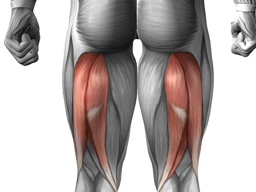
If hip pain is caused by an injury and is accompanied by dangerous signs, then you should immediately contact a traumatologist.
- Joint deformity.
- Inability to move your leg or hip.
- Inability to transfer weight to the affected leg.
- Strong pain.
- Sudden swelling.
- Any signs of infection (fever, chills, redness).
Biceps tendon rupture
The biceps femoris (anatomy, especially clinical, clearly shows this lesion) can suffer from rupture. It occurs when a muscle is severely stretched and partially pulled away from its junction with the bone. This injury requires long-term treatment and recovery.
When a rupture occurs, there is a loss of muscle strength, severe pain. The patient may report stabbing pain in the back of the thigh, which often comes on suddenly during exercise, and a clicking or popping sensation. When straightening the leg, overcoming resistance, the pain increases, and walking is also difficult.
A return to normal activity occurs in the absence of pain during the full range of motion. This type of injury heals within 1 to 2 weeks.
Symptoms of a Tendon Detachment:
- Intense pain after sudden movement.
- Excruciating pain when flexing the hip and extending the knee.
- Swelling in the affected area.
- The appearance of hematomas.
- Weakness of the hamstring, that is, a decrease in the ability to flex the knee joint with resistance.
- A fractured bone fragment can be felt through the skin in the event of an avulsion fracture.
Treatment for an avulsion of the biceps femoris tendon includes rest, ice application, compression, and lifting, which should be used immediately.
- Compression with an elastic bandage should be used for support.
- Consultation with a traumatologist is required.
- Strengthening and proprioception (kinesthesia) exercises are helpful.
- Deep tissue massage is beneficial for improving muscle elasticity.
- X-rays and ultrasounds are used to confirm the diagnosis of an avulsion fracture.
- For severe injuries or an avulsion fracture, surgery may be required to reattach the tendon and bone fragment.
- A comprehensive rehabilitation program should be started.
- After surgery, the knee should be kept at a right angle with a knee brace for several weeks.
Recovery time after surgery depends on the extent of the injury.
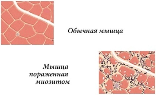
Tendinitis
The inflammation of the tendon tissue can begin as a result of frequent minor injuries. Cryotherapy and nonsteroidal anti-inflammatory drugs are used to treat inflammation.
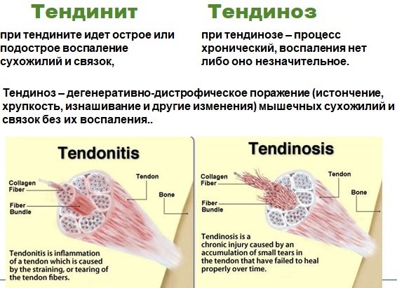
You can also apply bandages, heat, and electrotherapy. In addition, early physical therapy, as well as stretching and light training, are helpful in preventing loss of mobility and strength. Differential diagnosis should include runner's knee (Tibial Ilium Abrasion Syndrome) and damage to the external meniscus.
Symptoms:
- Soreness where the tendon meets the bone.
- Swelling at the junction of the tendon with the bone.
- If a person has tendinopathy of the biceps femoris, then they will have soreness in the outer or back of the knee. If tendons other than the biceps femoris are affected, soreness occurs medially.
- Pain with extreme flexion of the knee.
- Stiff knees after strenuous exercise or exercise.
- The tension in the muscles of the back of the thigh leads to limitation of its flexion.
Treatment:
- A good rest is required.
- The use of cold therapy.
- Consultation with a sports injury specialist.
- Anti-inflammatory drugs can be used to reduce pain, swelling, and inflammation.
- Ultrasound and laser will prove to be helpful.
- Conducting a comprehensive rehabilitation program with stretching and muscle strengthening exercises.
- Femoral massage.
Spasm
Cramps are painful muscle cramps that may occur after intense exercise or competition, dehydration, taking diuretics, or developing an imbalance in the minerals calcium, magnesium and potassium. A spasm occurs in any group of the biceps. Seizures are usually not dangerous, but can be very painful.
For spasms, the following can help:
- Stretching and massage. Stretch the spasmodic muscle and rub it gently until it relaxes.
- Try to pull the top of the foot on the affected side towards the head while the leg remains in a straight position. It helps relieve cramping in the hind thigh (hamstring). If you have a cramp in your front thigh (quadriceps), take a chair and try to pull your leg on the affected side towards your buttock.
- Applying heat or cold. You can apply a warm towel or heating pad to tight muscles. A warm bath or directing a hot shower onto spasmodic muscles can also help. You can relieve pain by massaging the affected area with ice.
The lower limb works as a unit, performing many repetitive tasks at home, at work, and during sports. Injuries to one area of the musculature, such as the biceps femoris, often indicate that anatomically other muscles have been damaged as well.
Author: Belyaeva Anna
Video about the biceps femoris
An overview of the posterior thigh muscle group:


