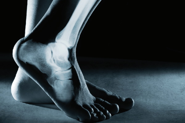
Unique ankle structure in the human body, it allows performing numerous complexly coordinated movements, due to where it is located and how it is mated with the knee and hip joint.
Among all musculoskeletal injuries, people often injure the ankle joint and ankle. Traumatologist is responsible for diagnostics and therapy. Correctly selected treatment will help prevent unpleasant consequences.
Record content:
- 1 What is an ankle, where is it located and what is it responsible for?
- 2 Features of the anatomy of the ankle joint
- 3 Ankle functions
- 4 Blood supply and nerve endings
- 5 Muscle
- 6 Ankle ligaments
- 7 Bone structures
- 8 Achilles tendon
- 9 Types of injuries
- 10 Diseases and their symptoms
- 11 Diagnostics
- 12 How are ankle diseases treated?
- 13 Ankle videos
What is an ankle, where is it located and what is it responsible for?
The ankle is the most important element that connects the bones of the lower leg and foot. Performs numerous functions. If it is damaged, a person's usual way of life is significantly disrupted, since he cannot move normally.
The upper border of the ankle joint is 5-7 cm higher from the medial ankle. The lower border is located at the junction of the two ankles. Everything below is related to the foot.
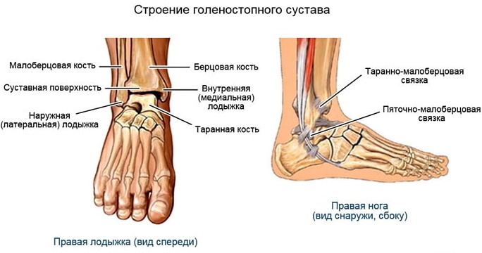
Features of the anatomy of the ankle joint
The ankle consists of 3 large parts:
| Name | Description |
| External | In this area are the joints, thanks to which the person wiggles the toes. Soft tissues are located on the sides, they strengthen the capsule, creating a movable connection. The outer part of the ankle allows a certain level of stability to be achieved. |
| Average | There are 2 main joints in this area: the calcaneo-cuboid and the talocalcaneal-navicular articulation. The latter makes more movements. |
| Back | The talus and heel bones are located in this area. They are responsible for depreciation. The back of the ankle is the strongest, it can support up to 350 kg of weight. If it is damaged, the person loses the ability to move the foot. |
The ankle structure also provides bone structures, ligaments and muscles that support the free movement of the foot. Normal blood circulation is carried out by blood vessels and arteries.
Ankle functions
The ankle is where the shin and foot meet. The joint in the human body plays an important role. If it is damaged, movement is impaired, and the ability to work is reduced. In some situations, the victim remains disabled.
The ankle joint performs the following functions:
- connects the bones of the lower leg and foot, ensuring their mobility;
- maintains the weight of the human body;
- evenly distributes the load on the foot;
- performs the functions of an upright position;
- allows a person to run, jump;
- makes it possible to make rotational movements with the foot, while the lower limb remains in an unchanged position.
The ankle joint also has a shock-absorbing effect. The impact force is softened when a person runs, walks or jumps. Helps to stand up straight and maintain balance while withstanding various physical activities.
Blood supply and nerve endings
Normal blood circulation is essential for the ankle to function properly.
The blood supply to this area is carried out by the following arteries:
- posterior fibular;
- anterior fibular;
- tibial.
The ramifications of the blood vessels pass through the ankle and wrap around the ankle joint. Blood enters the internal and external vessels (saphenous and tibial veins).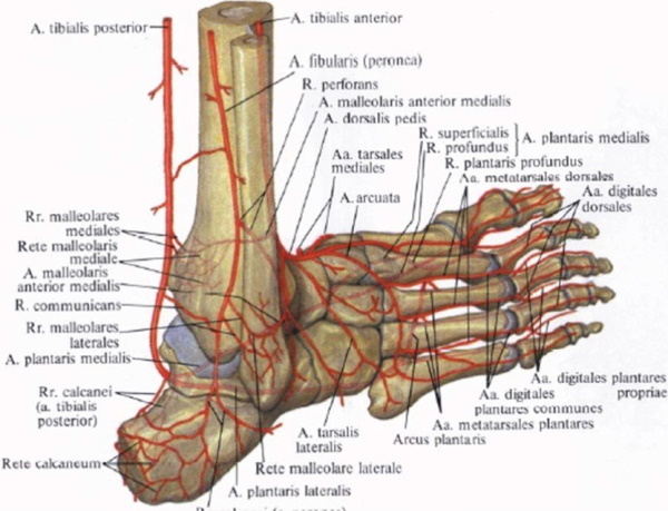
There are also a large number of nerve endings (gastrocnemius, peroneal, superficial and tibial). The branching of the nerve bundle in all adjacent areas occurs under the ankle, where the talus is located.
The most susceptible are blood vessels and nerve endings, which are located on the outside of the ankle. As a result of any injury, their integrity is violated.
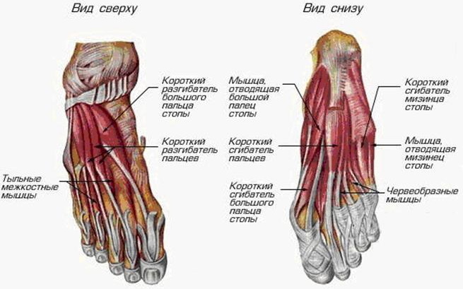
Muscle
The muscles in the ankle joint help maintain joint mobility, extension, flexion, and rotation. The musculature is located in front and behind in relation to the joint.
The ankle is surrounded by the following muscle groups:
| Name | Description |
| Bending | This group includes the musculature that helps to flex the toes. This also includes the plantar, triceps and tibialis posterior muscles. |
| Unbending | The muscles that take part in the extension of the fingers on the lower limb and the anterior tibial muscle. |
| Muscles for rotation | This group includes the long and short peroneal muscle. |
| Extension thumb muscles | We are talking about the peroneal anterior muscle (instep support). |
The well-coordinated functioning of all muscles allows a person to move around and perform various leg movements. In the case of the development of pathological processes, this ability is impaired and the risk of injury increases.
Ankle ligaments
Ligaments and tendons in the ankle area support the bones. Thanks to them, the functioning of the joint is controlled.
The following ligaments are located in the ankle area:
| Name | Description |
| Interosseous ligament | The joints that are located between the tibia. |
| Deltoid ligament (medial) | Supports the joint, preventing its collapse. |
| Tibial ligament | She controls the rotational movement of the foot. The ligament is located on the back of the ankle. |
The joint is also provided with a heel tendon, which gives it strength and allows it to withstand heavy loads.
Bone structures
The ankle joint consists of two main bones: the tibia and the tibia. The heel bone joins them. In the lower part of the ankle, a nest is formed, where the talus enters and a movable platform is formed.
Bone structures support the correct functioning of the ankle, and also take on a certain load. Thanks to them, a person makes jumps, steps, turns. Over time, bone structures wear out, resulting in an increased risk of injury.
The ankle joint contains the following bones:
| Name | Description |
| Ram | The bone consists of a body and a head, which are connected to each other by an expanded neck. The upper region (articular surface) is a necessary part that serves to connect with other bones. In the lower part of the talus structure, there is a groove that separates the joint. |
| Calcaneal | The largest bone in this part of the leg, which is flattened and oblong. Through the articular surface it connects to the talus and cuboid bone. |
| Scaphoid | The bone is located next to the inner edge of the foot. Helps to determine the height of the vault, easily probing under the skin. |
| Cuboid | The bone is located at the outer edge of the foot and connects to almost all elements of the ankle joint. |
| Wedge-shaped | There are several such bones and they make up the anterior tarsal region. |
The inside of the bones is covered with a thin layer of cartilage. It not only helps the entire ankle to function properly, but also reduces the stress on the bone structures. Damage to the articular cartilage gradually impairs the functioning of the ankle, against the background of which pathological processes (arthrosis, arthritis) develop over time.
Achilles tendon
The ankle is located in the area that connects the bones of the lower leg and foot. Its structure provides for such a complex element as the Achilles tendon, which is attached to the calcaneal tuberosity. Thanks to this complex structure, a person can stand or jump on one leg, rise on toes.
The Achilles tendon consists of the following muscles: gastrocnemius and soleus. They create an oval, inside which there is a hole, and around the lateral ankle and tendon muscle.
Types of injuries
The ankle joint is the most vulnerable in the human body. Active people who constantly move or play sports professionally face various injuries and injuries.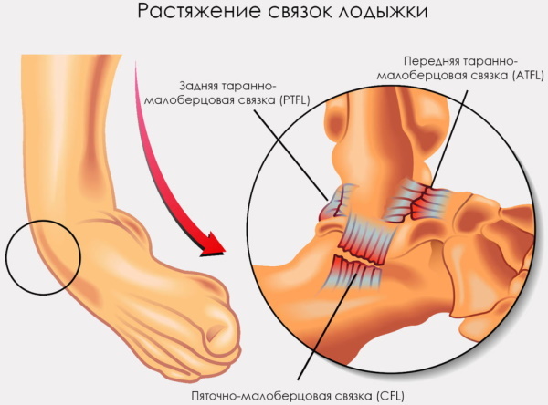
There are the following most common pathological conditions:
| Name | Description |
| Dislocation | Ligaments are damaged, acute painful sensations appear and soft tissues swell. After injury, hematomas, abrasions and swelling are formed. |
| Subluxation | Trauma often occurs in people with an incompetent ligament system. Repeated trauma negatively affects the functioning of the cartilage, provoking the development of arthrosis. |
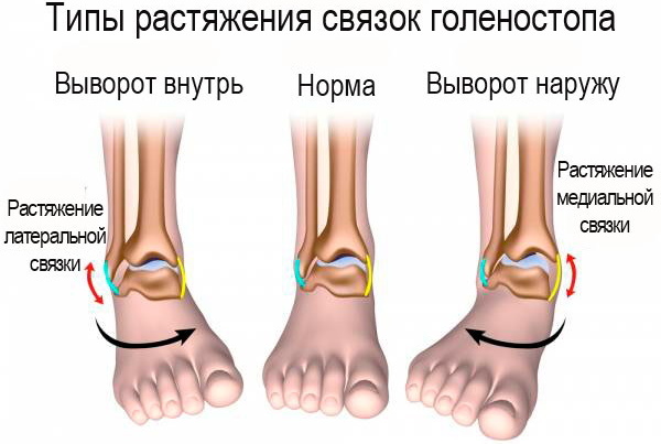
| Stretching | Damage occurs as a result of a sharp change in body position, when all the load falls on one leg. For this reason, partial or complete rupture of the ligaments can occur. |
| Injury | Injury that a person can receive while moving or in a collision, impact. The damage is accompanied by severe pain syndrome. A person cannot stand on his feet normally and correctly. Soft tissues swell, abrasions appear on the skin. |
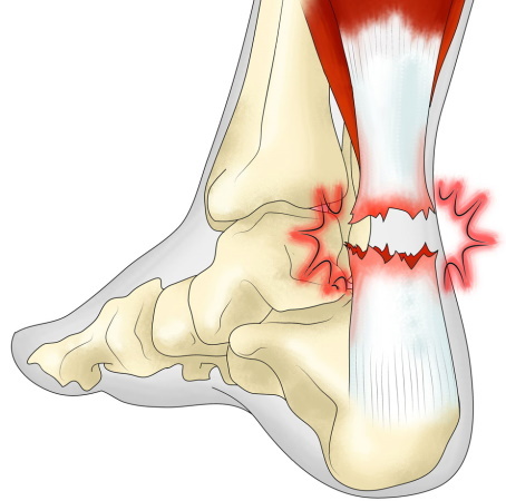
| Fracture | Trauma, after which it is impossible to get on the leg. When damaged, a crunch is heard and a sharp pain appears. Treatment in this case takes a long time and can lead to serious complications, since the injury occurs in the bone. |
A traumatologist will help to determine the type of damage and choose the most effective treatment. It is important to immediately go to the hospital, since improper therapy or its complete absence can provoke serious consequences, including disability.
Diseases and their symptoms
The ankle is located in such a place that the cause of damage can be not only physical influences, but also pathological processes occurring inside the human body:
| Name | Description |
| Deforming arthrosis | A disease that often occurs after an injury. In some situations, the cause of the pathology is a hereditary predisposition, extra pounds, and heavy physical exertion. Arthrosis is characterized by deformation of the surface of the bone structure, which leads to impaired smoothness of movements. |
| Arthritis | A pathological condition accompanied by an inflammatory process in the ankle joint. Soft tissues swell, the skin in the affected area turns red. As the disease progresses, fluid or pus accumulates in the joint area. |
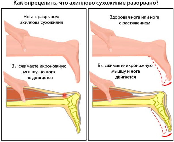
| Achilles tendon rupture | The main cause of the pathological condition is trauma. At the moment of damage to the Achilles tendon, a characteristic click appears. There is also a strong pain syndrome during movement. |
Pathological processes develop under the influence of certain factors, including the inflammatory process, mechanical influence, infectious sources and oncological neoplasms.
Diagnostics
A comprehensive examination of an ankle injury is necessary for the patient in order to make an accurate diagnosis and differentiate pathological processes. Considering the complaints of the person, the traumatologist prescribes the following diagnostic measures for the patient:
| Name | Description |
| X-ray | An affordable diagnostic method that is carried out in several projections. With its help, a specialist detects a fracture, dislocation, tumors or other pathological processes in the ankle area. |
| Magnetic resonance imaging (MRI) | The most informative diagnostic method that allows you to identify even minor injuries of the ankle joint. |
| Computed tomography (CT) | Diagnostics is assigned to patients if there are contraindications to MRI. The examination allows you to view bone structures and cartilage in order to identify neoplasms or arthrosis. |
| Ultrasound examination (ultrasound) | The examination will help identify pathological processes in the soft tissue area, view the joint cavity. |
| Arthroscopy | Minimally invasive diagnostic method. During medical manipulations, a camera is inserted into the joint cavity, which allows the doctor to visually examine the surrounding tissues of the ankle and identify pathological foci. |
In some situations, given medical reasons, a person is given a stress X-ray. The patient stands in a certain position, adhering to the required angle of inclination of the lower leg.
Additionally, patients are also prescribed laboratory tests (general blood count, urine test). The information received after diagnosis will allow the doctor to determine the disease and choose the most effective therapy.
How are ankle diseases treated?
The ankle is treated depending on the injury or illness that caused the limb to be affected. The doctor takes into account the results of a comprehensive examination, the condition of a person and the individual characteristics of his body, as well as where the pathological focus is located.
In the absence of visible damage to the skin and bones, the patient is provided with first aid:
- The victim is laid on a flat surface, providing him with complete rest.
- It is necessary to apply a cold compress to the damaged area.
- It is also important to restrict the movement of the injured joint. Apply a splint, bandage, taping, or an elastic bandage.
While an ambulance is being provided to a person, it is necessary to call a doctor.
The specialist will conduct an examination, prescribe a diagnosis and, based on the test results, select drug therapy for the victim:
| Drug group | Name | Application |
| Anti-inflammatory drugs | Diclofenac, Ketoprofen | Intramuscular injections are prescribed for adult patients. The dosage of the drug is 75 mg 1-2 times a day. The course of treatment lasts 4-5 days. |
| Muscle relaxants | Midocalm, Sirdalud | Medicines relieve muscle spasm and relieve pain. The adult dosage is 50-150 mg 3 times a day. The drug is administered intravenously or intramuscularly. In tablets, the medicine should be taken orally with meals. |
| Angioprotectors | Trental, Theonicol | Medicines improve blood circulation in the damaged area. Adult patients are prescribed 2-4 tablets 2-3 times a day. The drug should be swallowed whole, not chewed and washed down with plenty of water. |
| Chondroprotectors | Teraflex, Chondroitin | Medicines eliminate the symptoms of pathological processes and contribute to the restoration of cartilage tissue. The tablets are taken after meals, washed down with a glass of water. The recommended dosage for adults is 2 capsules 3 times a day. The course of treatment is determined by the doctor individually for each patient. |
| Means for external use | Ortofen, Finalgel | It is recommended to apply the gel or cream to the affected area of the leg 3-4 times a day for 2-4 g, rubbing gently with massage movements until the medicine is completely absorbed. |

Additionally, patients are prescribed vitamin and mineral complexes (Dekamevit, Pentovit). During the period of therapy, patients need to perform therapeutic exercises.
Specially selected exercises reduce swelling and help eliminate hematoma. The severity of pain syndrome also decreases, blood circulation in the area of the damaged ankle improves. Mobility is preserved, and metabolic processes are enhanced.
During the recovery period, to develop ankle mobility, patients are recommended to attend physiotherapy procedures:
- drug electrophoresis;
- ultrasound treatment;
- magnetic therapy;
- shock wave therapy.
In complex treatment, folk remedies can be used, but if there is no tendency to an allergic reaction. The components used can also provoke individual sensitivities. The ankle is an important joint in the human body. It is located in an area dominated by the complex structure of the lower limb.
Only a careful attitude towards it can prevent the occurrence of injury or pathological processes. In case of imminent damage, you must immediately go to the hospital. Timely medical attention will prevent serious complications.
Ankle videos
Ankle Anatomy: