
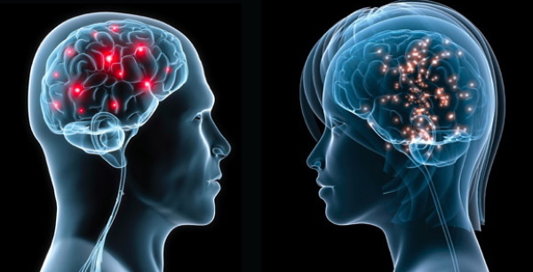
Cranial nerves control the functioning of internal organs, voluntary and involuntary movements of the body and face. The cranial nerves differ from the spinal nerves in anatomy, were formed later in the course of evolution.
With the development of human facial abilities and the appearance of speech, the need for careful innervation of the facial muscles to express emotions has increased. The brain plays a leading role, the spinal cord plays an executive role. It is also associated with the emergence of consciousness, motivation and the partial predominance of a person's volitional effort over instincts.
Record content:
- 1 Characteristic
- 2 Functions and properties
- 3 Anatomy and structure
-
4 Possible diseases and pathologies
-
4.1 Anosmia
- 4.1.1 Classification
- 4.1.2 Symptoms and Signs
- 4.1.3 Causes
- 4.1.4 Treatment methods
-
4.2 Bell's palsy
- 4.2.1 Classification
- 4.2.2 Symptoms and signs (external signs, how it manifests itself)
- 4.2.3 Causes
- 4.2.4 Treatment methods
-
4.3 Strabismus
- 4.3.1 Classification
- 4.3.2 Symptoms and Signs
- 4.3.3 Causes
- 4.3.4 Treatment methods
-
4.1 Anosmia
- 5 Cranial Nerve Videos
Characteristic
The cranial nerves (cranial nerves) are part of the human nervous system. Nerves are made up of nerve fibers - processes of neurons or axons. In turn, a neuron is a nerve cell, the smallest structural unit of the nervous system. The electrical impulse resulting from excitation moves along the axon of the neuron to the next cell.
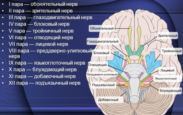
Thus, the nerves transmit impulses to each other and to other cells that are responsible for performing the corresponding functions. The function of FMN is the transmission of impulses from the brain to organs and muscles. There are 12 pairs of cranial (or cranial) nerves that perform their specific functions.
Each cranial nerve has a nucleus or several nuclei that process incoming information and make the appropriate decision. Depending on the number of nuclei and their functions, the nerve performs a particular task. The cranial nerves simplify the body's work, leading to the automation of the regulation of homeostasis (metabolism) and unconscious movements.
This is an important function that relieves stress from the brain. A modern person receives too much information, therefore, a conscious observation of the internal environment of the body and the simplest movements would lead to stress and overwork.
Functions and properties
Each pair of cranial nerves is functionally different from the others. The cranial nerve nucleus can perform 1 to 3 functions. Moreover, one nerve can both perceive information from receptors and carry out commands from the brain. The nerve is the link between the command organ and the performers.
The cranial nerves, the anatomy of which involves spreading throughout the body, provide the innervation of the organs. Innervation (organ control) is carried out by transmitting impulses from the brain to the corresponding organ for a specific task.
| Name | Serial number | Functions |
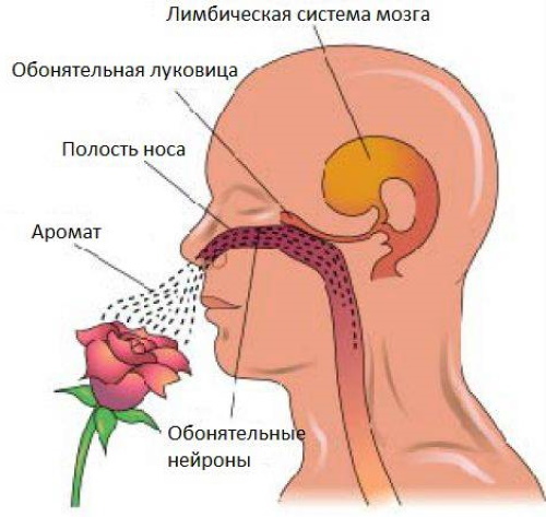
| Olfactory | I | Perception of surrounding odors, movement of impulses to the olfactory center. |
| Visual | II | Visual perception, transmission of images to the brain. |
| Oculomotor | III | Movement of the eyes, eyelids, reaction to light. |
| Block | IV | Pupil movement to the lower outer side. |
| Trigeminal | V | Sensitivity of the facial, oral and pharyngeal regions. The work of the muscles that provide chewing. |
| Diverting | VI | Pulls the eyeball outward. Controls just one muscle. |
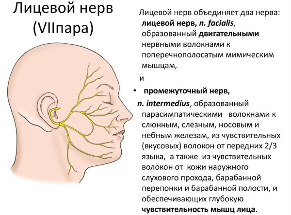
| Facial | Vii | It controls the facial muscles and the lacrimal gland, provides the blink reflex. |
| Vestibular cochlear or auditory | VIII | Conducts auditory impulses, maintains balance. |
| Glossopharyngeal | IX | Controls the muscle that lifts the pharynx. Responsible for the secretion of the gland near the ear. Provides sensitivity to the areas of the pharynx, partially soft palate, pharyngeal tonsils, the posterior third of the tongue, responsible for the perception of the bitter taste and taste of the imago. |
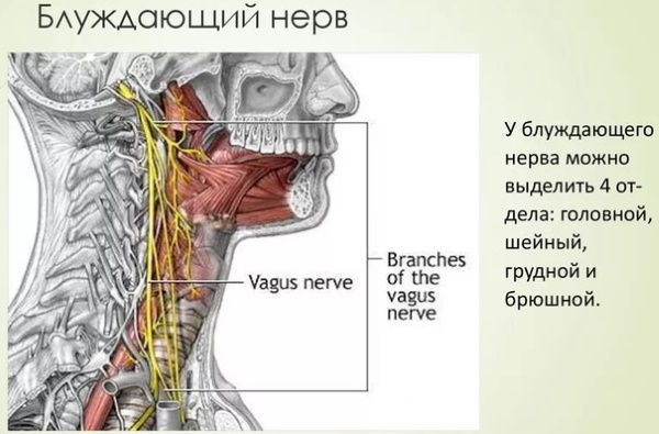
| Wandering | X | Movement of the muscles of the palate, pharynx, stomach, lungs and esophagus. Affects the production of secretions from the glands of the pancreas and stomach. Partially provides sensitivity to the pharynx and larynx, ear. |
| Additional | XI | Controls specific movements of the shoulder girdle and head. Often these are reflex involuntary movements. |
| Sublingual | XII | Innervates the muscles of the tongue. Provides voluntary and involuntary movements of the tongue. Participates in the act of sucking, licking, chewing and swallowing. |
The block nerve is responsible for only 1 action. The ability to quickly look away was evolutionarily important for humans. With the help of the block nerve, the individual managed to quickly turn his gaze to a potential source of danger.
The most versatile cranial nerve is the vagus nerve. It innervates almost all parts of the body.
Anatomy and structure
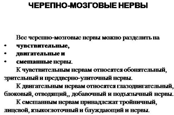
The cranial nerves, whose anatomy is highly specific, move from the brain, have afferent (ascending) and efferent (descending) pathways. Afferent pathways begin in the organs and ascend to the brain. Efferent impulses conduct impulses from the brain - the governing organ, to the executive organs. The performers can be glands, muscles, and receptors.
The glands produce certain substances: hormones, fluids. Muscles contract or relax. Receptors perceive the environment and the internal environment of the body. Separate external receptors, for example, located on the skin and perceiving the environment, and internal - determining the composition of the blood, the fullness of the stomach.
The olfactory and optic nerves carry only afferent (ascending to the brain) information from receptors, so it cannot be said that they “leave” the brain.
Structure:
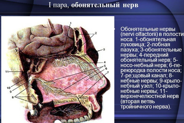
- The pathway of the olfactory nerve begins with the nasopharyngeal mucosa. There are receptors responsible for smell recognition. On the mucous membrane, there are olfactory networks of 15-20 nerves, which are collected higher in the olfactory bulb. Then the impulses are sent to the olfactory triangle and there they are divided into two olfactory strips. The collected olfactory information reaches the corresponding center in the temporal lobes of the cerebral hemispheres and is processed.
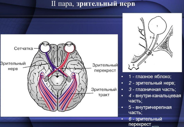
- Like the olfactory nerve, the optic nerve is predominantly ascending. With the help of sight, the individual receives 90% of the information about the world around him. The optic nerve begins in the nerve cells of the retina. Then the impulses are collected in the bundle of the optic nerve, pass through the orbit and are sent to the temporal lobes of the brain. The visual path ends in the visual zone of the brain - the occipital lobe.
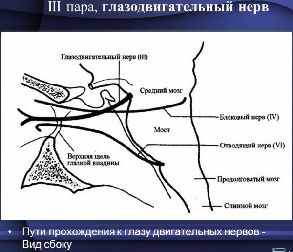
- The cranial oculomotor nerve is mixed. There are both ascending and descending paths in its anatomy. The efferent pathway begins in the cerebral peduncle, passes between the posterior and superior cerebellar artery, outside the internal carotid artery, and ends in the orbital cavity. Before entering the orbit, the nerve is divided into an upper and lower branch. The first is responsible for raising the century. The second is divided into 3 more branches and innervates the lower rectus, lower oblique muscles of the eye. These muscles are responsible for vertical movement of the eyeball.
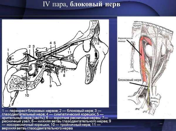
- The block nerve innervates only one muscle. The path begins at the level of the midbrain, bends around the brain stem and exits the gap between the temporal lobe of the cerebral hemisphere and the leg. The legs of the brain are paired structures of the lower part of the brain. They are mostly conductors of upward and downward impulses, are involved in information processing. Having overcome the gap near the temporal lobe, the trochlear nerve penetrates the meninges and goes into the orbit.
- The descending pathways of the trigeminal nerves leave the pons varoli and are sent to the temporal bone. The Varoliev bridge is a brain structure that connects the spinal cord and brain through the cranial nerves. He is responsible for processing incoming information and provides the brain with "feedback" - a report on the work performed by the organs. In the temporal bone, the trigeminal tract is divided into the optic, superior and delicate nerves. They provide tactile sensitivity of the face and mouth, and are responsible for the chewing movements of the jaw.
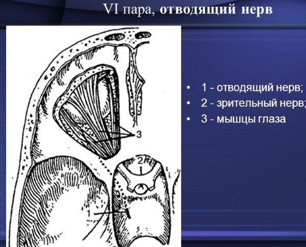
- The abducens nerve has only efferent (descending) pathways. This is the motor nerve. Overcoming the thickness of the brain, the nerve passes near the internal carotid artery and enters the orbit. It innervates the muscle that pulls the eyeball outward.
- The facial nerve is predominantly a motor, that is, descending. Due to the fact that the intermediate nerve is attached to it, the facial one also has afferent pathways. The cranial nerve exits through the auditory opening into the parotid gland. There it is divided into 5 branches. They form the eardrum, the string that provides auditory perception. Before leaving the temporal bone, the facial nerve forms 4 more branches. All of these branches are found on the face and temples. Descending, the nerve encircles the neck, cheekbones, lower jaw and lip. The facial nerve controls virtually all of the muscles in the face. Thus, the facial nerve innervates almost all the muscles of the face.
- The vestibular cochlear nerve conducts impulses from the vestibular apparatus and the ear. That is, this cranial nerve is responsible for spatial orientation and hearing. It has a vestibular and auditory root, which transmit relevant information. The path lies in the brain and near the cochlea.
- Glossopharyngeal nerve from the brain is directed to the temporal bone. Further down, near the internal carotid artery. It enters the root of the tongue and gives off 2 branches: the tympanic nerve and glossopharyngeal nerves, which are also divided into 5 branches. The tympanic nerve is sensitive and is involved in the transmission of auditory information. Glossopharyngeal nerves extend from the trunk of the main nerve and innervate the muscles of the mouth. The lingual branches penetrate the tongue and provide gustatory sensitivity.
- The vagus nerve has the head, cervical, thoracic, and abdominal regions. The vagus pathway begins in the brain, down to the neck and chest. In the cervical region, the vagus nerve splits into the pharyngeal, laryngeal and cervical branches. Further, in the thoracic region - on the thoracic, bronchial branches and the pulmonary, esophageal plexus. In the abdominal region, it is divided into the posterior and anterior trunk, which innervate the gastric muscles. Thus, the vagus nerve controls almost all voluntary movements. Its lesions can cause serious pathologies.
- The accessory nerve has a cerebral and spinal part. It only has descending paths. The pathways of the accessory nerve leave the brain and penetrate into the cranial cavity, dividing into two branches. The internal branch is part of the vagus nerve. The outer branch joins the trapezius muscle. The latter is located at the back, between the shoulder blades, and has the shape of a triangle.
- The hypoglossal nerve leaves the medulla oblongata and travels to the carotid artery. It enters the lingual muscles and divides into 4 branches. They innervate the lower surface of the tongue and its skeletal muscles, which provide the acts of swallowing, chewing, sucking and licking.
Possible diseases and pathologies
Damage to the cranial nerves causes serious abnormalities in the functioning of the body. Diagnosis of disorders of the FMN is carried out by means of a neurological examination.
Anosmia
Complete or partial loss of smell. Anosmic patients cannot perceive all odors or certain ones. Also, loss of smell can occur due to age.
Violation of the perception of smell does not allow you to fully feel the taste of food, emotionally suppresses the patient.
Classification
Divide congenital and acquired anosmia. Acquired anosmia can result from damage to the olfactory nerve. A congenital disease occurs due to pathologies at birth: a defect in the nasopharynx, mucous membrane. Cranial nerves, the anatomy of which is defective, can also be congenital anosmia.
Symptoms and Signs
The main symptom of anosmia is loss of sense of smell. Patients who notice this symptom should consult an ENT doctor. He prescribes appropriate diagnostics to identify the causes of the disease. Often, anosmia is diagnosed using olfactometry - a study in which a person needs to recognize the odorous substances presented by a specialist.
Causes
The reasons include:
- Diseases of the nasopharyngeal mucosa (colds, rhinitis).
- Traumatic brain injury.
- Smoking.
- Parkinson's disease.
- Alzheimer's disease.
- COVID-19.
- Polyp of the nose.
Treatment methods
If, after examining a specialist, it turned out that the cause is inflammation of the mucous membrane, then the following treatment methods are suitable. Camomile tea.
Recipe:
- 1 tsp steamed chamomile leaves with a glass of boiling water.

- Infuse the broth for 15 minutes. until partial cooling. The infusion should be warm while straining.
Application: wash the nasal cavity 2 times a day for 5-10 days. It is necessary to draw in the liquid through the nostrils and exhale through the nose or mouth. Collection of herbs.
Recipe:
- 30 g dried sage flowers, calendula;
- 20 g of dried herb St. John's wort, plantain;
- 10 g of field horsetail, spring primrose.
- Mix the ingredients for 1 tbsp. spoon 1 cup boiling water.
- Boil the mixture for 1 minute.
- Insist for 30-40 minutes and drain.
Application: charge a spray bottle with a decoction, inject into the nose and irrigate 2 times a day for 10 days. Saline solutions: Aquamaris, physiomer and aqualor help to cope with anosmia. Use according to the instructions for the preparation. Apply for 5-10 days. Aloe leaf drops.
Another effective remedy is aloe drops:
- Keep 1-3 aloe leaves in the refrigerator for 2 weeks.
- Squeeze out the juice, dilute with cold boiled water in a ratio of 1 to 10.
Application: drip into the nose up to 4 times a day, 4-6 drops in each nostril. The course lasts 5-10 days.
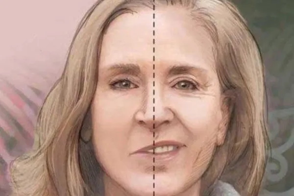
Bell's palsy
Cranial nerves whose anatomy is damaged can cause nerve palsy. Bell's palsy is a common form of a defect in the facial nerve. Every year new clinical manifestations of this disease are revealed. The incidence is approximately 23 people per 100,000 each year.
Bell's palsy has nothing to do with stroke, but they can be confused because the clinical manifestations are similar in some ways. In both stroke and paralysis, prompt medical attention and long-term treatment are needed. Modern medicine is able to fight Bell's palsy and quickly remove the consequences of the disease.
Classification
Paralysis can spread to one side of the face or, in rare cases, both.
There are 6 degrees of severity of the disease:
- No pathology. Muscles are normal.
- Presence of minor violations. At this stage, during movement, there is a noticeable asymmetry of the left and right sides of the face.
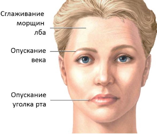
- Moderate pathology. Asymmetry is also observed in calmness. The affected eyebrow and the corner of the mouth are lowered, the eyes are completely closed.
- Moderate severity of pathology. The face is distorted, there is no symmetry. The eyelids do not drop completely, the muscles of the forehead are immobilized.
- Severe pathology. The muscles of the face do not contract. In tranquility, the left and right sides are asymmetrical.
- Complete paralysis. With maximum effort, the paralyzed muscles do not contract. The face is skewed, not asymmetrical.
Symptoms and signs (external signs, how it manifests itself)
Paralysis comes on suddenly. Symptoms include severe weakness, pain near the ear. In some patients, the perception of the taste of food disappears. Paralysis is cured in 80% of patients in 1-2 weeks or months.
Early symptoms include a slight increase in temperature, stiffness on one side of the face.
Causes
Paralysis can occur for a number of reasons. It includes both infectious diseases and injuries. Bell's palsy is often the result of an inflammation of the facial nerve.
The causes of the disease include the following:
- Hypothermia.
- Colds.
- In a certain season.
- Flu.
- Rubella.
- Diabetes mellitus.
- Tumors of the brain and parotid gland.
- High blood pressure.
- Injuries to the head and facial nerve.
Treatment methods
Ancillary treatments for Bell's palsy are:
- Acupuncture that relaxes muscles and relieves pain. It also helps guide nerve impulses along the facial nerve.
- Gymnastics. Prevention and rehabilitation method. Facial gymnastics includes smiling with a closed mouth, squinting, closing and opening the eyes, and raising the eyebrows.
- Traditional methods of prevention is treatment with mud or paraffin compresses. Their temperature should be between 40 and 500 degrees, depending on the type of compress.
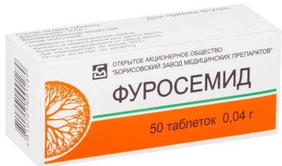
- Clinically, paralysis is treated with Furosemide 40 mg per day.
- Alpha lipoic acid is given. Recommended Allowance: 600 mg per day.
- Ipidacrine. Improves the conduction of nerve impulses. It is applied at 20 mg 3 times a day.
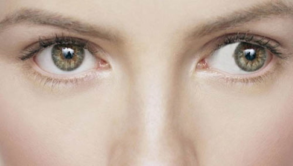
Strabismus
This is a disease that can be caused by abdominal nerve pathology. Strabismus is manifested in the deviation of the eyeball from the object in question. The eyes cannot be directed to one point.
Classification
Acquired and congenital, horizontal and vertical strabismus are divided.
More often diagnosed horizontally converging (to one point) or diverging (the eye is diverted to the other side) strabismus:
- Monocular strabismus involves abduction to the side of one eye, alternating - abduction of two eyes.
- Alternating strabismus differs from monocular in that a person looks at an object variably. Moreover, both eyes are used.
Symptoms and Signs
Symptoms of strabismus include:
- Deviation of the eye to the side.
- "Floating" look.
- Limited eye mobility.
- Lack of symmetry of the gaze.
- Dizziness.
- Head deviation.
Causes
The causes of strabismus can be congenital or acquired.
During the examination, the following causes of the disease can be identified:
- Injuries.
- Paralysis.
- Congenital pathologies.
- Prolonged stress.
- Influenza, COVID-19, other infectious diseases.
- Mental trauma, such as severe fright.
- Farsightedness or severe myopia.
Treatment methods
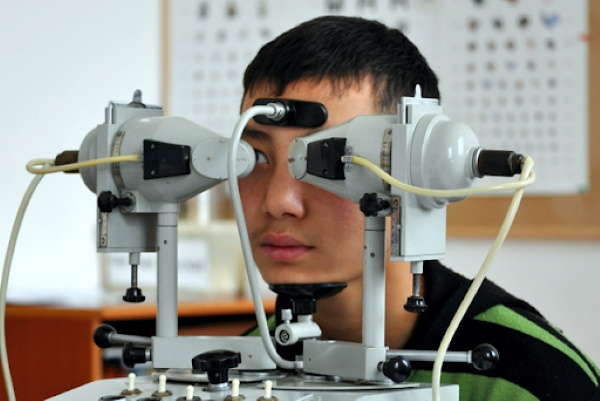
Strabismus is treated with medicines, computer programs, and surgery:
- Pleoptic way. Intentional severe stress on the defect eye. Treatment by means of special computer technologies.
- Orthopedic method. Special techniques and computer programs are used to restore the symmetry of the gaze.
- Diploptic method. The most gentle. Assumes natural eye strain in normal conditions.
-
Modern medicine offers the devices "Sinoptophor" and "Monobinoscope" for the treatment of strabismus. The number of procedures is prescribed individually by ophthalmologists.
- In surgery, strabismus is treated with botox. It is pricked in the oculomotor muscles. Thus, the affected muscles stop pulling the rest in their direction and the position of the eye is normalized. Possible adverse effects, such as a constant feeling of grit in the eyes. There is dryness, cutting pain when blinking.
When the cranial nerve is damaged, they try to improve its conduction, correct defects in anatomy. This is done through surgical intervention and preventive work with computers.
Cranial Nerve Videos
Cranial nerves - features:


