
Bone and muscle structures of the legs - one of the strongest in the human body. Anatomically, the lower limb refers to the portion of the leg from the knee to the ankle, known as the lower leg. A description and photographs of its structure are presented later in the article.
Record content:
- 1 Calf location, functions
-
2 The anatomical structure of the shin bones
- 2.1 Tibia
- 2.2 Fibula
- 3 Features of the articulation of the bones of the lower leg
- 4 Lower leg vessels
- 5 Calf nerves
-
6 Calf muscles. Their anatomy, classification and functions performed
- 6.1 Anterior muscles
- 6.2 Lateral muscles
- 6.3 Back muscles
- 7 Diseases associated with the lower leg, their symptoms and causes
- 8 Complications of diseases, possible consequences
- 9 Shin video
Calf location, functions
The lower leg together with the thigh forms the lower limb. It is located between the knee and ankle, and the upper leg is located between the thigh and knee.
Functionally, the calf muscle is necessary for everyday walking and being in an upright position, for more vigorous activity. It controls balance, movement of the foot and toes. It is the most active muscle when running, walking, jumping, and therefore the most commonly injured.
The main bones of the lower limb, tibia and fibula, connect the ankle to the knee. They work together to stabilize the ankle joint to provide support for the calf muscles, but the tibia supports a significant portion of the body's weight.
The anatomical structure of the shin bones
The shin of a human leg (the photo in the article illustrates bone formations) consists of 2 long bones, tibia and fibula, which are very strong skeletal structures:
- The tibia is located near the midline of the leg and is thicker and stronger. It connects to the thigh bone to form the knee
- The fibula is much smaller and located on the lateral (further from the midline) side. It is attached to the tibial under the knee joint and serves as an attachment point for the lower leg muscles. They are both tubular in structure, but unequal in size.
Tibia
The tibia (Tibia) is located in front of the lower leg, connecting the knee joint to the bones of the ankle.
It has a 3-sided shape and 3 distinct edges:
- proximal;
- rod;
- distal.
Tibia refers to the long bones that are found on the upper and lower limbs, fingers and toes. Long bones contain bone marrow in a cavity along the length of the rod.
At the ends of the tibia there is a cancellous bone, where pockets of blood circulation and bone marrow are located, which appear cancellous under a microscope. The entire length of the tibia is covered with a layer of compact substance, which gives it strength.
The upper part of the tibia forms part of the knee and is known as the tibial plateau, on which the femur rests and together they form the hinge of the knee.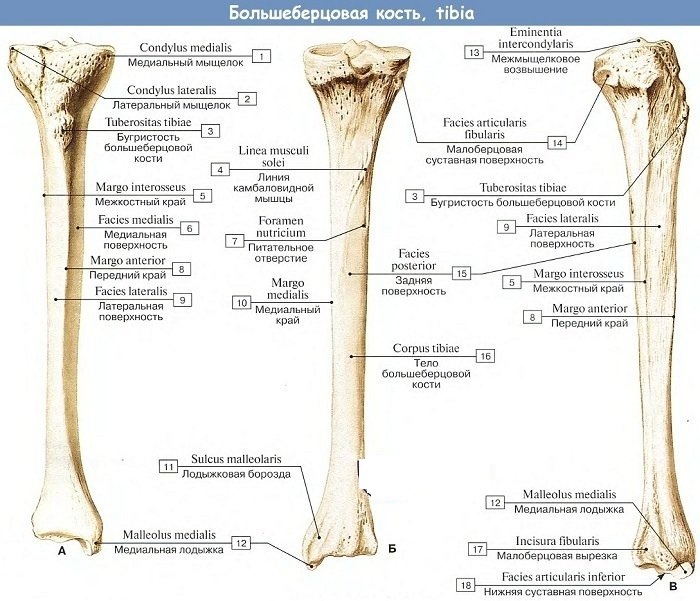
It contains two condyles, rounded projections that help the tibia to fit into the base of the femur. The lateral condyle is located above the head of the fibula, and the medial condyle is opposite. The front of the top of the tibia is called the tuberosity, where the patella attaches through the patellar ligament.
The lower part of the tibia rests on the apex and medial part of the talus. The medial portion of the tibia is called the medial malleolus.
The main function of the tibia is motor. Many of the basic human movements are based on the strength of the tibia. Since it forms the knee joint with the femur and the ankle joint with the tarsal bones and the fibula, it supports the movement of the muscles of the foot and ankle joint, which are involved in walking, running, jumping.
The tibia provides stability and stress to the lower leg. It gives the leg leverage, makes it easier to walk, run, climb, kick.
Fibula
The fibula, Fibula, is a thin, long bone that attaches near and slightly below the tibia. It has very little mass, provides lateral stability to the lower leg, and acts as a traction to increase the range of motion of the ankle, especially lateral and medial rotation of the foot.
It is the thinnest of the long bones. The word fibula in Latin means "brooch", and many believe that it is named so because anatomically connected to the tibia, it forms a safety pin reminiscent of an ancient brooch.
Long bones have a spongy end and dense bone along the shaft. Along the length of the rod, in the center of the fibula, there is a cavity filled with red bone marrow.
The separation of the cancellous and compact bones is the epiphyseal plate (growth plate). This is the site of growing tissue formation in children and adolescents. After the completion of bone growth, this area is replaced by bone tissue.
The middle fibula is about 390 mm long in adult males and about 360 mm in females.
Looking at the cross section of the bar, 3 different shapes can be seen:
- 3-sided;
- 4-sided;
- wrong.
Each fibula can have more than one type of cross-section, and the combinations differ between males and females. The fibula is the thinnest longest bone in the body in terms of width to length.
The fibula is located on the side (outside) of the tibia, slightly posteriorly (towards the back) and is displaced slightly lower. The proximal (upper) end of the fibula articulates with the lateral condyle of the tibia, just below the knee. This is called the proximal tibiofibular joint. The fibula is not part of the knee joint.
The distal (inferior) end of the fibula articulates with the tibia in a depression called the peroneal notch or distal tibiofibular joint.
Even more distally, the fibula articulates with the talus at the talofibula, which forms part of the ankle joint called the lateral malleolus. It can be felt on the outside as a hard protrusion on the outside of the ankle.
The fibula provides lateral stability to the lower limb and ankle. Its articulation with the tibia and talus provides additional range of motion during ankle rotation.
The fibula in a healthy person does not provide support for significant body weight.
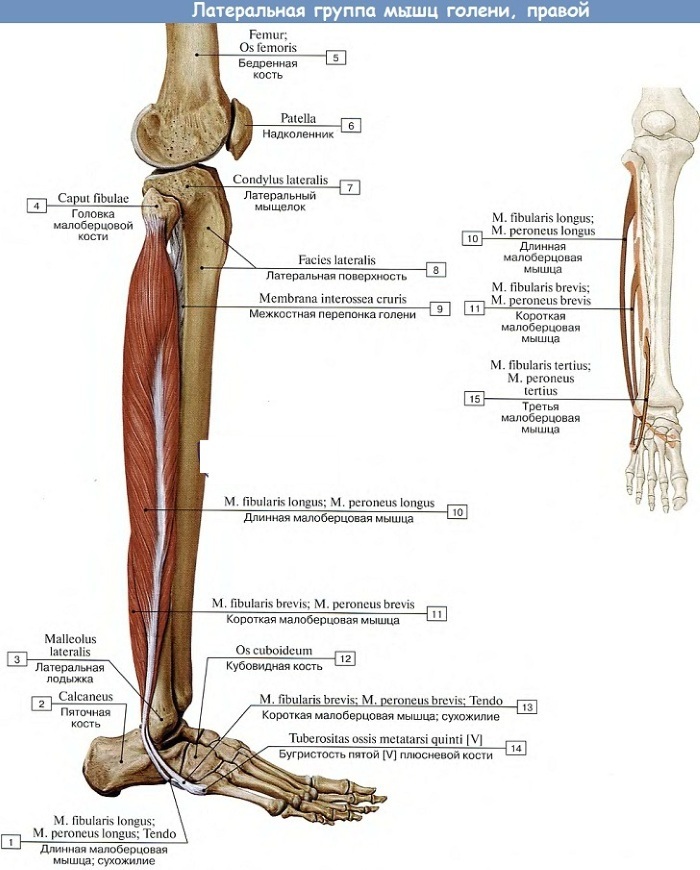
Features of the articulation of the bones of the lower leg
All muscle groups on the lower leg in front and behind are interconnected with the help of special connections. The tibia is connected to the fibula interosseous membrane.
The fibula connects to the tibia through a network of connective tissue that runs almost the entire length of the fibular shaft. The proximal tibiofibular joint is held in place with the lateral peroneal collateral ligament.
Lower leg vessels
The shin of the human leg (the photo can be clearly seen in the anatomical atlas) is supplied with blood due to the anterior tibial, posterior tibial and peroneal arteries.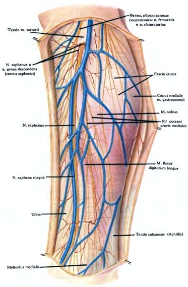
These blood vessels supply oxygen and nutrients to the surrounding structures:
- bones;
- muscles;
- nerves.
The tibia is supplied with blood through the nutritional artery and through the periosteal vessels from the anterior tibial artery.
Calf nerves
The lower leg of a person, and especially the foot, as can be seen in the photo, have a rather bizarre pattern of innervation of the skin by terminal branches.
For example, the skin of the foot is innervated by 7 separate nerves:
- Superficial peroneal nerve.
- Deep peroneal nerve.
- The sural nerve.
- Saphenous nerve.
- Calcaneal branch of the tibial nerve.
- The medial branch of the plantar nerve.
- Lateral branch of the plantar nerve.
There are 2 main nerves in the leg: the femoral one serves the front of the leg, and the sciatic one controls the back. The nerves of the legs can have many nerve roots, and when pain or discomfort is felt in these areas, it usually indicates a pinched or pinched nerve. By the location of the nerve pain, you can determine which of the nerves is damaged.
- The femoral nerve is the largest part of the lumbar plexus.
- The sciatic nerve is the longest and widest nerve in the body. It starts at the lower back and goes down to the lower leg. Along the way through the legs, the sciatic nerve splits into the tibial and common peroneal nerves, which in turn split into many smaller nerves in the legs and feet.
The anterior tibia is innervated by the deep peroneal nerve, the lateral by the superficial peroneal nerve, and the posterior by the tibial nerve.
Calf muscles. Their anatomy, classification and functions performed
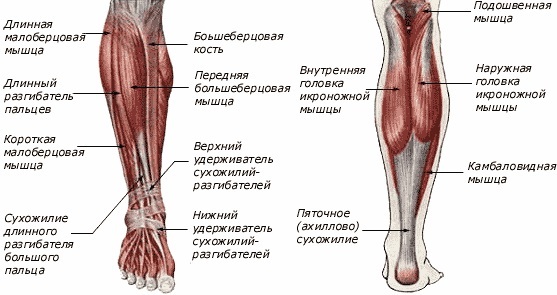
The shin of a human leg (a photo of the muscle structure is presented later in the article) is represented by several muscle groups. The main muscle is the calf, which gives the calf a muscular appearance. The muscles of the tibia are found on the front and back of the lower leg.
The muscles related to the fibula are located on the front of the lower leg. The Achilles tendon, or Achilles, attaches the muscles of the calves to the bones of the ankle and foot.
Anterior muscles
The tibialis anterior muscle is a muscle located in the front of the lower leg. Muscles run from the area just below the knee, down the front of the lower leg, and attach to the top of the foot. The anterior muscles allow dorsiflexion and help guide the toes upward.
The anterior tibial muscle helps to lift the ankle and foot off the ground, helps to tighten the leg, rearrange. Since the tibialis anterior muscle attaches to the top of the foot, it helps in the arch of the foot.
Lateral muscles
The lateral muscles are located on the outside of the lower leg.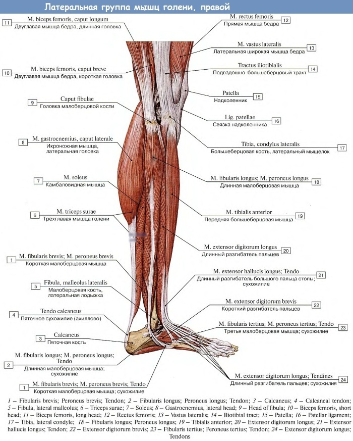
 These are short and long peroneal muscles that help to straighten or bend the foot, to raise its lateral edge.
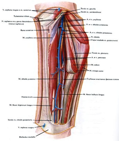
Back muscles
The posterior section contains large muscles:
- gastrocnemius;
- soleus;
- plantar.
The calf is shorter, thicker, and under it lies the soleus. The Achilles tendon serves as the attachment point for these 3 muscles. Their function is to flex the sole.
Inside the back of the lower leg is a deep posterior compartment. Here is the tibialis posterior muscle, the long flexor of the fingers and the separate long flexor of the big toe. The hind tibial gives the foot rigidity, and the long flexors are responsible for their parts: the big toe and the rest of the toes. All three help with plantar flexion.
Diseases associated with the lower leg, their symptoms and causes
The shin of a human leg (the photo is presented in the article for informational purposes) consists of bones, blood vessels, muscles, connective tissue. They are important for movement and standing. Sports, jogging, falling, or an accident can injure your feet. Common injuries include sprains, joint dislocations, and bone fractures. They can affect the entire leg or only the foot, ankle, knee, or hip.
Certain medical conditions also lead to foot problems. For example, knee osteoarthritis, which is common in older people, can cause pain and limited movement. Vein problems lead to varicose veins or deep vein thrombosis.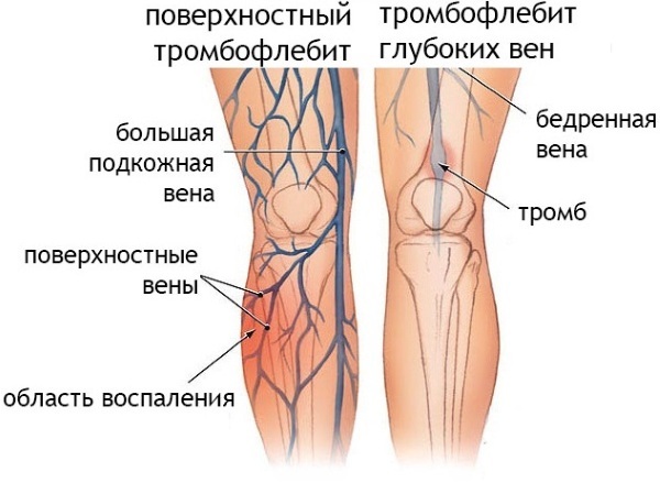
| Diseases | Causes | Symptoms |
| Peripheral artery disease | In this condition, the limbs, usually the legs, do not receive enough blood. This is often due to narrowing of the arteries. | Feel weakness in the legs, numbness, or cramps when walking. They can be cold and unusual in color. |
| Deep vein thrombosis | A blood clot formed in a vein, usually in the thigh or lower leg. | This does not always cause symptoms, but there may be pain, swelling in the leg, and the skin may be warm and red. |
| Peripheral neuropathy | This happens when the nerves in the body are damaged, which carry messages to and from the brain. Diabetes is the most common cause, but other illnesses, medications, injuries, or infections can cause it. | If it affects the nerves in the legs, they feel tingling, numb, or weak. |
| Electrolyte imbalance | Electrolytes are minerals like sodium, potassium, and calcium that help muscles to function properly. | With the loss of a large amount of fluid in the legs, you can feel compression, numbness, weakness. |
| Spinal stenosis | This condition occurs when the gaps in the bones of the spine are narrowed. | The condition puts pressure on the nerves in that area and can cause pain, tingling, numbness, or weakness in the legs. Also, the balance when walking can be imbalanced. |
| Radiculitis | Leg pain that occurs due to a pinched nerve in the lower spine, displacement of the intervertebral disc or hernia, displacement of the vertebra, spasm of the gluteal muscles, or spinal stenosis. | The pain can range from severe cramps to severe shooting pains that make it difficult to stand or sit. |
| Crick | The muscle is stretched too much during sports, when walking. | The pain is severe and begins immediately, and the affected area becomes tender to the touch. |
| Sprain | Trauma occurs when the tissue that connects the bones stretches or breaks. Ankle sprains are common. | The injured area is swollen and sore, and weight cannot be transferred to it. |
| Muscle cramps | Occurs when a muscle is suddenly compressed, usually in the calf. Seizures tend to occur with age, and they can also occur if a person is outside for a long time in hot weather and does not drink enough water. Spasms usually go away on their own and are not a sign of any health problem. | This can cause severe pain, and the muscle under the skin becomes hard to the touch. |
| Medial Tibial Stress Syndrome | It happens when the muscles and tissues around the shinbone become inflamed, causing the inner edge of the bone to hurt. They are common among people who run a lot. Flat feet, stiff arches, or improper shoes can also lead to them. | It is best to rest your legs, apply cold compresses for 20 minutes several times a day, and take pain relievers. |
| Crack in the bone | If the stress-like pain persists, there may be a small fissure in the shinbone. This happens when the muscles around the bone are overloaded and do not cushion the impact of the movement as they should. | Rest is the best treatment for a stress fracture, but it can take 6 to 8 weeks to heal. |
| Tendinitis | Tendons are flexible cords that connect muscles to bones. If they become inflamed, it can be very damaging, especially when a person moves this joint. This is called tendinitis, and it is a wear-and-tear injury that can affect your hip, knee, or ankle. | One of the first warning signs of inflammation is pain in the lower leg. |
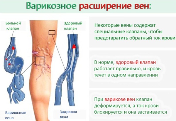
| Phlebeurysm | When the veins have to work hard to get blood back to the heart, they swell and look twisted, blue or deep purple. | Swollen veins cause a feeling of heaviness in the legs, burning, throbbing, or cramps. |
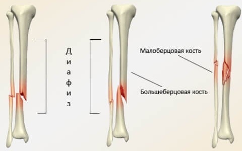
| Fractures of the shin bones | Ankle inversion with significant stress. Clumsy landing after falling. Direct injury to the anterior or inner leg or ankle. A sport involving a sudden change of direction, like football. |
Instant pain. Swelling appears immediately. Inability to walk and pain when walking. Soreness in the leg. Bruising may be present. The patient cannot bear the load on the leg. The leg is crooked or deformed. |
Medical treatment can help for fractures, leg cramps, blood clots, or nerve problems. In other cases, it is recommended to use compresses, rest the feet, and wear appropriate shoes.
Complications of diseases, possible consequences
The most common complications occur after injuries, fractures:
- Pain in knees or ankles.
- Bone infection (osteomyelitis).
- Damage to a nerve or blood vessel.
- Compartmental syndrome or neuromuscular disease.
- Arthritis.
- Unequal leg length. A baby's long bones grow from the ends of the bones in softer areas called growth plates. If the fracture passes through the growth plate, that limb may eventually become shorter or longer than the opposite limb.
Deep vein thrombosis can lead to a serious condition called pulmonary embolism, when a blood clot breaks off and travels to the lungs.
Legs are the basis of the whole body, a person actively moves with their help. The lower leg and ankle should be trained to withstand significant forces and be more resistant to injury. In the photographs, you can see the main muscles that help keep the body in good shape, and make walking controlled.
Shin video
Calf muscles 3D overview:


