The cerebral hemispheres are difficult organized nerve center. The histological structure of the cortex is represented by various layers, in which there are up to 16 billion. neurons.
Record content:
- 1 Characteristic
-
2 Functions
- 2.1 Frontal lobe
- 2.2 Parietal lobe
- 2.3 Occipital lobe
- 2.4 Temporal lobe
-
3 Neurons
- 3.1 Pyramidal
- 3.2 Star-shaped
- 3.3 Fusiform
- 3.4 Other types of cells
-
4 Layers
- 4.1 Supragranular layers
- 4.2 Inner granular layer
- 4.3 Infragranular layers
- 5 Cytoarchitectonic fields
-
6 Nerve fibers
- 6.1 Associative
- 6.2 Projection
- 6.3 Commissural
- 7 Modular organization principle
- 8 Possible diseases and pathologies
- 9 Video about the cerebral cortex
Characteristic
The image of the brain is familiar to every person - it is a structure in the shape of a helmet, the surface of which, intersected by convolutions and grooves, has a characteristic wrinkle.
The cerebral cortex is the outermost layer of nerve tissue that makes a person unique. The hemispheres are focused on the regulation of voluntary actions, sensory processing, learning, and memory.
Certain areas of the brain receive information directly from sensory inputs - eyes, nose, skin, mouth, ears, muscles, organs - but they can also be modulated by memory and emotional centers brain. This was made possible thanks to the convolutions and deep grooves.
A large surface is concentrated in a small space, like a crumpled towel is a gimmick evolution, which allows, with a limited volume of the skull, to provide the brain with enough space to solve various tasks. The minimized size of the skull is associated with the limited width of the female birth canal.
The evolutionary process led to the fact that over time, the human cerebral cortex underwent a process corticalization or wrinkling, which is associated with the vast knowledge that the human brain has accumulated over the course time.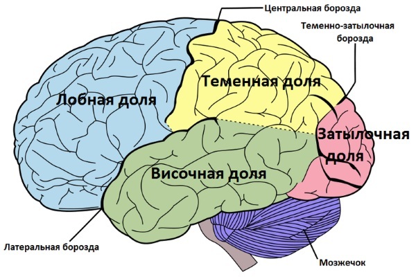
The outer layer of the brain is made up of gray matter, which contains neurons. Gray matter is like a "generator" in which current (pulses) is generated and then transmitted to other parts of the central nervous system or body.
The cerebral cortex (the histology of the brain is presented in some detail in the medical atlas) is divided into 4 lobes, each of which performs a specific function.
Functions
The cerebral cortex is only the outermost layer of the gray matter, where most of the actual information processing takes place. The hemispheres cover sensory and motor areas. The former receive data from the thalamus and process information related to the senses.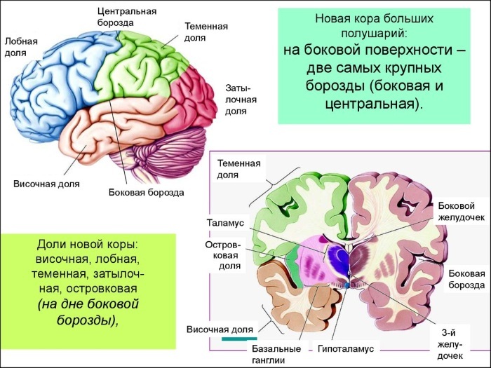
Within sensory zones there are associative zones that give meaning to sensations and associate them with specific actions. The cerebral cortex is divided into smaller areas structurally (grooves) and histologically by cellular organization (layers). The latter leads to the appearance of Brodmann zones. Together, this information provides insight into the functions of areas of the brain.
The most important ones are:
| Frontal lobe | The place where "decisions are made." Plays an important role in planning, decision making and movement, including speech. |
| Parietal lobe | Responsible for somatic sensory processing or feelings. |
| Temporal lobe | Responsible for processing the auditory senses (touch and hearing). |
| Occipital lobe | Responsible for processing visual information (vision). |
| Special zones of Brodmann | Wernicke's zone is responsible for the fluency of speech. Broca's zone - for motor speech. |
The cerebral cortex is the youngest layer, but also the most complex, where the highest analysis and synthesis of all incoming information takes place.
Frontal lobe
It is the part of the brain that controls important human cognitive skills such as emotional expression, problem solving, memory, language, judgment and sexual behavior. In fact, it is a "control panel" of personality and communication skills.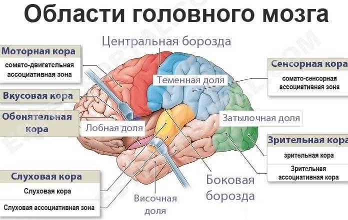
It is also responsible for primary motor function, or the ability to consciously move muscles, and for two key areas associated with speech, including Broca's area.
Parietal lobe
This is the area where impulses from the skin such as heat, cold, pain and touch are interpreted. As with the primary motor area in the frontal lobe, the more sensory information comes from areas of the body, for example, from the fingers, the greater the surface area of the parietal lobe is involved in the processing of this information.
The parietal lobe is also an important piece of spatial information that makes it possible to judge size, distance and shape. A special triangular area, the parietal associative cortex, makes it possible to understand written language and solve mathematical problems.
Occipital lobe
The occipital cortex is located at the back of the skull. One of the most important parts of this lobe is the primary visual cortex, which receives data from the retina. This is where the mind interprets color and other important aspects of vision.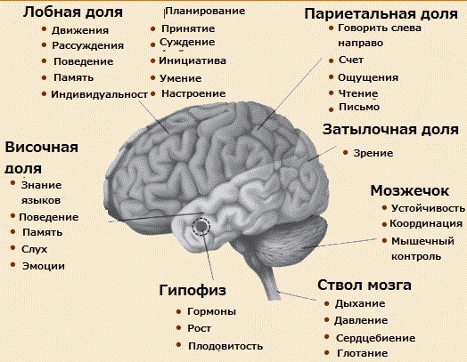
The occipital lobe contains various areas related to visual communication. One area is the place of obtaining visual images of the language (area of visual perception), and the other is the place of their interpretation (area of visual associations).
This is important for reading and reading comprehension. For example, if a person sees words in another language, but does not understand that language, he will only use the visual reception area.
Temporal lobe
The temporal lobes of the cortex are located on either side of the head, flush with the ears.
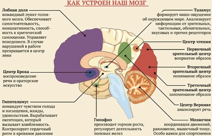
They coordinate specific functions, including visual memory (recognizing faces), verbal memory (understanding language), and interpreting the emotions and reactions of others.
Neurons
The cerebral cortex (histology and the functional significance of neurons are under constant study) consists of a huge number neurons, and they all represent different variations of 3 morphological forms: pyramidal, fusiform and stellate (granular cells).
By function and structure, neurons are represented by 60 types (candelabrum cell, with a double bouquet of dendrites, basket, columnar basket, and many others). In shape, they are only pyramidal and not pyramidal. Other cell types seen in the cortex are modifications of one of 3 morphological species.
Pyramidal
The characteristic neuron of the gray matter of the cerebral cortex is the pyramidal cell, which gets its name from its triangular cell body. These neurons make up up to 75% of the cellular component of the cortex and are the main types of neurons.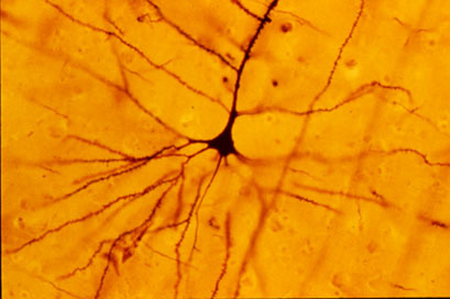
They vary in size and can be small or gigantic. These cells have a thick branching dendrite (branching like tree branches) that extends to the surface of the bark, and several basal (underlying) dendrites.
The number of basal dendrites varies widely, but there are usually more than 3-4 primary dendrites that branch off to the next generations of dendrites (secondary, tertiary).
They all branch out near the cell body. On the opposite side of the cell, an axon (neurite, a long, cylindrical process) descends from the thick dendrite, which exits the cortex and enters the subcortical white matter. The axon is difficult to see and may not be present in all cells.
Both small and large pyramidal cells mainly perform activities related to movement.
Star-shaped
Stellate (granular) cells are usually small, and because their processes are projected in all planes, they resemble a star. They are located throughout the bark, with the exception of the very surface layer. Their processes are very short and protrude locally on the surface of the brain.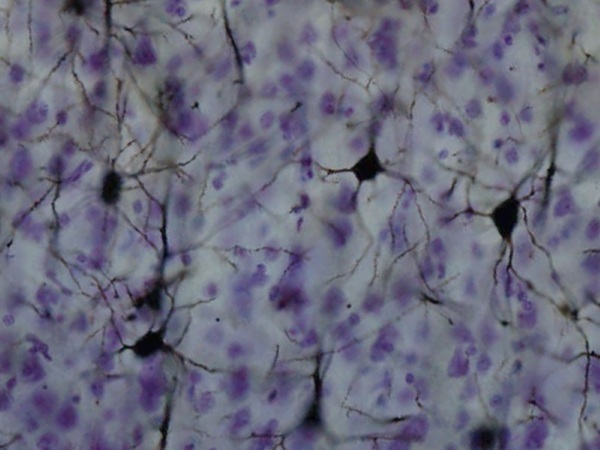
They are able to inhibit or activate the energy of other neurons in the cortex. Depending on whether their dendrites have spines (small cytoplasmic protrusions), they are subdivided into spine and aspen cells (without dendritic spines).
Spines are mainly located in layer IV, where they secrete glutamate, which is an excitatory neurotransmitter, so they are functionally excitatory interneurons. Aspinates release gamma-aminobutyric acid (GABA), which is the most potent inhibitory neurotransmitter in the central nervous system, so they act as inhibitory interneurons.
Fusiform
Fusiform cells are usually located in the deepest cortical layer. Their dendrites are projected onto the surface of the cortex, and their long axons are directed vertically or horizontally.
Among neurons, one can distinguish commissural cells (whose axons go to the other hemisphere), associative (whose axons connect areas of the cortex within one hemisphere) or projection oriented (axons directed to the lower parts of the brain).
Other types of cells
Cajal cells, small, spindle-shaped, horizontally spaced neurons, are found in the outermost layer of the cerebral cortex. They are very rare and only a few of them can be found on the surface of the adult brain. These cells are able to synthesize and secrete the glycoprotein reelin, a protein found in the brain and responsible for the memory and learning mechanisms.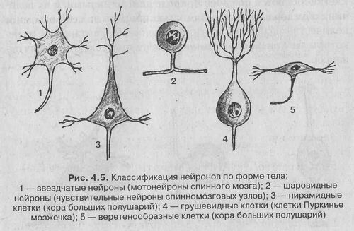
Martinotti cells are multipolar neurons that are most densely located in the deepest layer of the cortex. Their numerous axons and dendrites point towards the surface of the cortex, where they branch out into the molecular layer.
Layers
The cerebral cortex, the histology of which is constantly being examined, is built according to the screen type. This means that neurons are structurally and functionally arranged in layers or plates. The six layers of this part of the cortex are numbered in Roman numerals, and each cortical (outer) layer contains neurons of different shapes, sizes, and densities, as well as different nerve fiber organizations.
Functionally, the layers can be divided into 3 parts.
Supragranular layers
The group includes layers that are above the level of layer IV: the molecular plate (layer I), in which there are few neurons, the outer granular (layer II), the outer pyramidal plate (layer III).
They are the completion of intracortical connections, which are either associative (associated with other areas of the same hemisphere), or commissural (having connections with the opposite hemisphere, primarily through the corpus callosum body).
Inner granular layer
Layer IV, granular, inner, consists mainly of stellate neurons. The cells in this layer send their dendrites to different layers of the cortex, especially the molecular layer, while their axons move deeper into the cortex.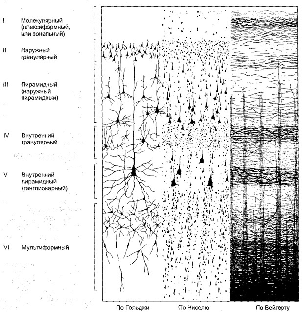
In addition to the intracortical synapse, the axons of this layer can be long enough to form associative fibers that pass through the white matter and end up in various structures of the central nervous system.
Infragranular layers
The group includes layers below layer IV: layer V - the inner pyramidal plate and layer VI - a multimorphic plate, they primarily connect the cerebral cortex with the subcortical areas. These layers are most developed in the motor areas of the cortex.
The motor areas have very small granular layers and are often referred to as "agranular cortex". Layer V gives rise to all major cortical efferent (motor) projections of the basal ganglia of the brainstem and spinal cord. Layer VI projects primarily onto the thalamus.
Cytoarchitectonic fields
The histology of the brain, which received its true development only after the creation of a light microscope, determined the differences in the parts of the gray matter in the cerebral cortex. Since the human brain contains a large number of neurons, it is definitely one of the most complex structures in the human body.
Not only the type of nerve cells, but also their relationship is heterogeneous in different parts of the cortex. The specificity of the organization of neurons is called cytoarchitectonics. The cerebral cortex map was created in the late 19th and early 20th centuries. German neurologist K. Brodman identified 52 cytoarchitectonic fields of the cerebral cortex.
This is a complex nervous mechanism, consisting of receptors, conductors of the brain center and nerve impulses, in which all stimuli emanating from the environment and the body are analyzed and activated person.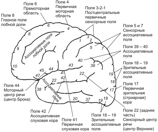
Examining the anatomical and genetically the cerebral cortex, scientists have compiled a cytoarchitectonic map of the brain.
In the process of evolution, genetically separate 5 zones of the crust were formed:
- neocortex;
- archicortex;
- paleocortex;
- periarchicortex;
- peripaleocortex.
The very first occupies 95% of the area of the entire surface of the cerebral hemispheres. Its structure is layered, consisting of 6 layers. The new cortex is also subdivided into areas: occipital, temporal, parietal, frontal. Each of the areas, in turn, is divided into fields, which, according to K. Brodman, more than 50.
Nerve fibers
In each cell layer, in addition to nerve and glial cells, there are nerve fibers - outgrowths of cells of a given layer or parts of the brain.The structure and density of fibers are not the same in different parts of the cortex. The fibrous structure of the cortex corresponds to its cellular composition.
The cerebral cortex contains a huge number of cellular bodies, from which fibers of varying complexity extend. They are divided into 3 categories.
Associative
Bundles of nerve fibers that connect one area in the cerebral cortex to another area in the same hemisphere. Associative nerve fibers emerging from the cerebral cortex are located in one hemisphere and connect different functional centers. Long fibers connect different lobes of the hemisphere, and short associative fibers connect the convolutions within one half.
Projection
Nerve fibers carry impulses to and from the cortex. Some projection paths of fibers connect the cerebral cortex with the brain stem, diencephalon, spinal cord. Others carry signals up the cerebral cortex. 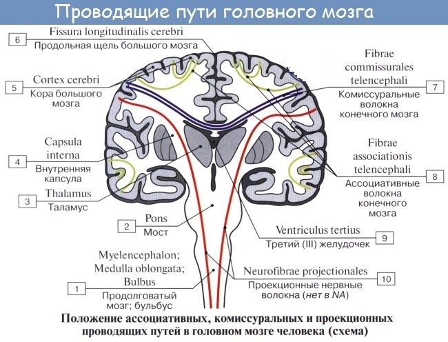 Such fibrous tracts form a wide layer around the thalamus and basal nuclei. It is an internal capsule that subsequently fan out into specific areas of the cortex.
Commissural
Nerve or commissural fibers that connect one hemisphere to the other. The largest junction is the corpus callosum, which consists of about 300 thousand cubic meters. fibers. Damage to this pathway can cause a split brain in some people, in which a person can literally feel like they have two minds.
The anterior commissure is a smaller connection between the hemispheres that connects the structures of the anterior temporal lobe, including the amygdala and other structures of the olfactory lobe.
Modular organization principle
The cerebral cortex has not only horizontal structures, but also vertical formations called columns or columns. In fact, they are functional units of the cerebral cortex or module. Each column is oriented perpendicular to the surface of the cortex and consists of all 6 cell layers.
Neurons are closely connected within a single column, although they share common connections with adjacent and distant columns, as well as with subcortical structures, especially the thalamus. These columns are capable of memorizing relationships and performing more complex operations than a single neuron.
The module has 3 departments:
- The entrance, which is formed by fibers that carry information from the visual and other parts of the cortex.
- Informative processing area. It is a system of interconnected stellate and pyramidal neurons.
- An exit consisting of pyramidal neuron axons that transmit nerve impulses outside the column.
Modules are able to select an action depending on the current state and the nature of the input signals they receive. This ability is essential for correct problem solving. The process of solving problems in columns can be compared to the same process in artificial intelligence.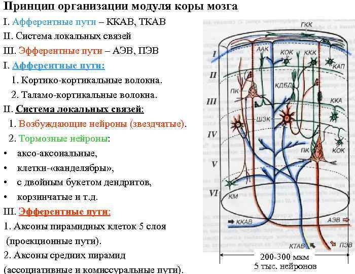
Thus, each column should:
- Set a goal and initiate the state necessary to achieve this goal
- Define a set of possible actions that allow you to move from one state to another.
- Determine intermediate subgoals (points that need to be achieved in the process of achieving the final goal)
- Finally, reach the goal from the initial state by completing intermediate subgoals, each time approaching the final goal.
This means that at any given time, the cerebral cortex can cope with many problems, while simultaneously posing multiple targets and defining multiple initial states with simultaneous and interdependent activation of different amounts cortical pillars.
Possible diseases and pathologies
The cerebral cortex (histology reveals various pathologies) is rarely affected in isolation. Symptoms of the disease are usually associated with the brain and occur simultaneously with damage to the white matter of the hemispheres.
The largest damage to the cerebral cortex is accompanied by the disappearance of mental activity.
A number of disorders occur as a result of damage or death of the brain cells of the cortex. Symptoms experienced depend on the affected area.
- Apraxia Is a group of disorders characterized by the inability to perform certain motor tasks, although there is no damage to the motor or sensory function of the nerves. People may have difficulty walking and may not be able to dress or use normal objects properly. Apraxia is common in people with Alzheimer's disease, parkinsonism, and diseases of the frontal lobe.
- Agrafia, this is damage to the parietal lobe of the cerebral cortex. At the same time, people have difficulty writing or cannot write at all.
- Ataxia. Damage to the cerebral cortex can also lead to a lack of muscle control or coordination of voluntary movements, such as walking or lifting objects. Ataxia, which is a symptom of an underlying disorder, can interfere with various movements and make speech, eye movement, and swallowing difficulties.
Injuries in this area of the brain contribute to the development of depressive disorders, difficulty making decisions, lack of control over impulses, problems with memory and attention.
The cerebral cortex is the outer layer of the cerebral hemispheres. It is divided into areas with specific functions such as sight, hearing, smell, and sensation, and controls higher mental processes such as speech, thinking, and memory.
The study of morphology, histological studies help to understand how different parts of the brain function, as well as to comprehend how a disease or injury affects certain functions.
Video about the cerebral cortex
The cerebral cortex: