
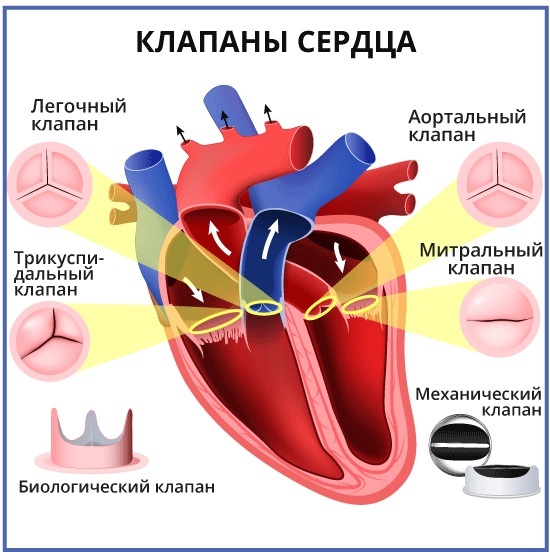
Any changes in the work of the cardiovascular system are dangerous to human life. Impaired blood circulation provokes numerous diseases and complications. One of the important elements of the functioning of the heart is tricuspid valve, which is located on the right side of the organ.
His defeat provokes the appearance of various signs, with which it is important to go to the hospital in a timely manner. Correct diagnosis and treatment regimen will help prevent the consequences.
Record content:
- 1 Structure, localization, anatomy of the tricuspid heart valve
- 2 Organ work
- 3 Valve functions
- 4 Normal Valve Performance
- 5 Pathology of the 3-leaf valve of the heart, causes and signs
- 6 Dysfunction stages
- 7 Diagnostic methods
- 8 Methods for treating diseases
- 9 Indications and types of operations
- 10 Prognosis for pathologies
- 11 Valve Disease Videos
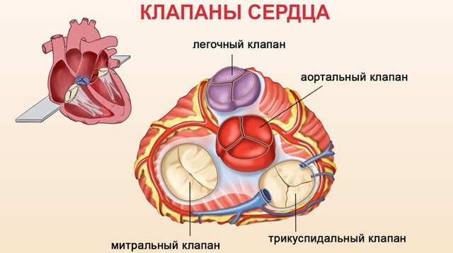
Structure, localization, anatomy of the tricuspid heart valve
The tricuspid valve of the heart is located in the atrium and ventricle on the right side.
Its structure is represented by the following elements:
| Name | Description |
| Fibrous ring | Consists of a large number of elastic fibers. Attaches to the atrial septum. A large number of cardiac conduction channels pass through the annulus fibrosus. Some of this element is represented by muscle fibers and has a loose structure. As the annulus fibrosus moves away from the atrial septum, its thickness gradually becomes smaller. |
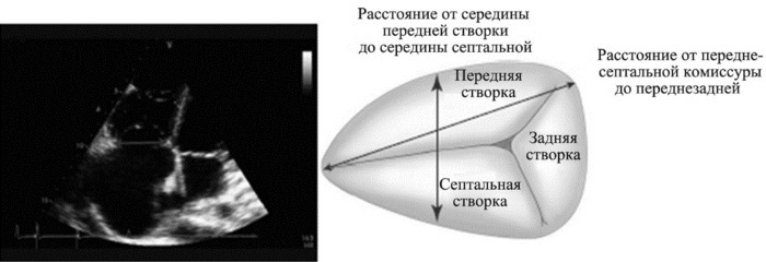
| Sash | In most cases, the normal development of the heart valve involves 3 main leaflets (anterior, posterior, and septal). Sometimes their number increases, according to experts, due to the bifurcation of the posterior flap. They all vary in size. The largest flap is the main one. The structure of these elements provides for the presence of a base, a body and a closing section. |
| Tendon chords | There are 3 main elements, each of which performs its own specific functions. |
| Papillary musculature | The muscle is located on top of the atrial septum. Tendon chords extend from it. |
The condition and function of the tricuspid valve is important for normal circulation. It prevents backflow of blood into the right atrium.
Organ work
The function of the tricuspid heart valve is to maintain normal circulation throughout the body. Passing through all cells and internal organs, blood gives off oxygen, and back receives carbon dioxide. It is also saturated with decay products, due to which it acquires a dark color.
The work of the tricuspid valve is necessary to separate the jointly functioning elements (left and right ventricles, atria). Venous blood through these sections returns to the pulmonary circulation. It is saturated with oxygen and through the left atrium, the ventricle flows into the systemic circulation.
Valve functions
The tricuspid valve of the heart is located on its right side and is an important element in the work of the pulmonary circulation.
It performs the following functions:
- With heartbeats, it prevents the blood flow from moving from the lower part of the right ventricle to the atrium.
- Takes a direct part in the process of blood circulation. The tricuspid valve is responsible for the delivery of venous blood to the vessels that are located in the bronchi.
- Supports gas exchange in the alveoli of the lung tissue and heat transfer.
Continuous blood circulation in the human body throughout his life is responsible for the rapid and timely delivery of saturated blood with oxygen to all internal organs and tissues. Through the venous outflow, carbon dioxide and decay products leave the body. In medicine, this mechanism is called the cardiovascular system.
The systemic circulation begins from the left ventricle, and ends in the right atrium. The small circle of blood flow is directed from the right ventricle to the left atrium.
Normal Valve Performance
The tricuspid valve of the heart is located between the right ventricle and the atrium. When the heart muscle relaxes, it opens up. When the myocardium contracts, the valve closes. The valves are tightly closed due to the presence of chords and muscles. During normal operation of the tricuspid valve, venous blood flows from the right heart to the left. Then it is sent to the lungs for gas exchange.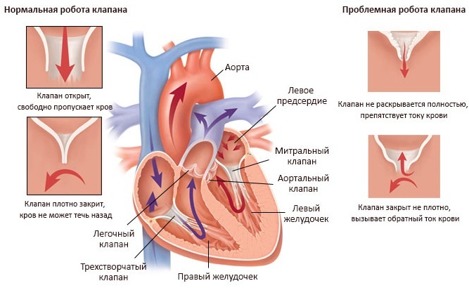
Reverse flow of blood into the cardiac cavity from large vessels is not observed due to the correct functioning of this valve. With the development of pathological processes, the entire mechanism is disturbed, as evidenced by the emerging clinical symptoms.
Pathology of the 3-leaf valve of the heart, causes and signs
In most cases, pathological changes in the work of the tricuspid valve provoke heart failure or the development of stenosis. Degenerative processes disrupt blood circulation, resulting in characteristic clinical signs.
| Name | Description |
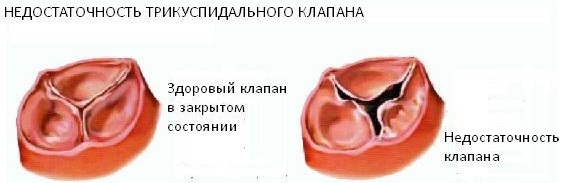
| Failure | A disease characterized by insufficient closure of the tricuspid valve. In such a situation, the blood returns back to the atrium. |
| Stenosis | The pathological condition is characterized by a narrowing of the holes that are located in the tricuspid valve of the heart. In most cases, problems arise against the background of rheumatism, congenital defects or after prolonged mechanical impact on the chest. |
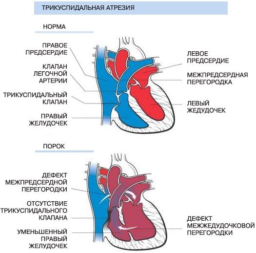
| Congenital malformations | Pathological changes occur during the period of intrauterine development of the human body, often against the background of hereditary diseases in the mother's body. |
Pathological processes in the area of the tricuspid heart valve develop as a result of the following provoking reasons:
- complication of rheumatism;
- severe thyrotoxicosis;
- the development of myocarditis;
- infective endocarditis;
- cardiomyopathy;
- carcinoid syndrome;
- mechanical trauma to the chest, which resulted in damage to the heart;
- surgical intervention in the area of the mitral valve, due to which its leaflets have increased;
- myocardial infarction;
- an increase in the annulus fibrosus in diameter.
Congenital defects in most cases develop together with other hereditary abnormalities (open oval window, atrial septal defect).
Dysfunction of the tricuspid valve is also provoked by tumor neoplasms that have appeared on various internal organs. In this situation, disruption of the functioning of the cardiovascular system occurs as a result of the toxic effects of malignant processes.
Developing pathological changes, given the main reason for their appearance, provoke the emergence of certain clinical signs. Symptoms will help the cardiologist determine the degree of damage to the cardiovascular system and assess its ability to function.
Dysfunction of the tricuspid valve is accompanied by the following symptoms:
- the shade of the skin on the face changes;
- shortness of breath appears;
- breathing is disturbed;
- swelling of the face;
- there is a general weakness in the body;
- the patient is worried about vomiting, the secreted masses contain blood impurities;
- metabolism in the intestinal area is disturbed;
- the person gets tired quickly;
- painful sensations appear in the area on the right side under the ribs.
 The liver also enlarges, not only the face swells, but also the lower, upper limbs. Excess fluid builds up in the lungs. The intensity of the clinical picture and the presence of certain signs depends on the degree of development of pathological processes.
It is important, if any symptom appears, to go to the hospital and undergo a complete examination in order to establish an accurate diagnosis. Correctly selected therapy will prevent serious consequences.
Dysfunction stages
The tricuspid valve of the heart is located on the right side of the organ. Pathological changes develop gradually. In medicine, disorders of the functioning of the tricuspid valve are distinguished, given the complexity of pathological changes and the possibility of their reversal.
There are the following stages in the development of organ damage:
| Name | Description |
| Stage I | At this stage of the pathological processes, minor disturbances occur that affect the movement of the small and large circulation. |
| Stage II | Pathological processes progress, but if treatment is started in a timely manner, degenerative changes can be reversed. |
| Stage III | Significant problems in the work of the tricuspid valve provoke the appearance of specific signs, for example, an increase in blood pressure. |
| Stage IV | At the last stage of the disease, the structure of the valve is affected. A person's condition worsens, progressive symptoms require urgent hospitalization and strict supervision of the attending physician. |
At the last stage of valve damage, pathological processes are irreversible. Correctly selected therapy will improve the patient's quality of life and prevent complications.
Diagnostic methods
A comprehensive examination will help the doctor establish the correct diagnosis and choose the most effective treatment regimen.
Patients with lesions of the tricuspid heart valve are prescribed the following diagnostic methods: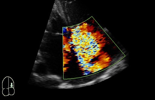
| Name | Description |
| Electrocardiogram (ECG) | An examination method that will assess the work of the heart and identify the pathological abnormalities present. Determine also the volumes of the atria. |
| Phonocardiogram | During the study, a specialist detects systolic murmurs. |
| X-ray examination | Based on the images, the doctor will determine the dilatation of the affected ventricle or atrium, the size of the organ itself, and identify congestion. |
| Echocardiography | The cardiac septa and their movements are seen. Deformation of the valves and the appearance of new formations are also revealed. |
| Spiral computed tomography (CT) | The specialist evaluates the functioning of the main muscle in the human body. |
| Catheterization | The diagnostic method is carried out to determine the pressure in the right heart. |
An important defining moment in the course of diagnosis is the systolic murmur during exhalation. Its presence indicates the development of tricuspid valve insufficiency. In addition, or before surgery, patients are prescribed coronocardiography. A diagnostic method that will assess blood circulation and detect pathological changes.
Methods for treating diseases
The tricuspid valve of the heart (located between the right atrium and the ventricle) is treated depending on the degree and stage of its defeat, how extensively pathological processes develop, their area localization. During the period of therapy, it is important to strictly follow all the doctor's recommendations, without exception, since the disease is dangerous to the patient's life.
Drug therapy for lesions of the tricuspid heart valve involves the use of the following drugs:
| Drug group | Name | Application |
| Diuretics | Britomar, Hydrochlorothiazide | Medicines reduce congestion in the patient's body and remove excess fluid. The tablets are taken orally, regardless of food, and washed down with a sufficient amount of water. Adult patients are prescribed 10-20 mg once a day. If necessary, the dosage is increased to 20-40 mg. |
| Potassium supplements | Panangin, Asparkam | The drugs prevent the accumulation of excess fluid in the patient's body. The medicine is taken orally 1-2 pills 3 times a day after meals. The minimum course of therapy lasts 2-3 weeks. In severe situations, the medicine is administered intravenously through a dropper. |
| Venous dilators | Korvaton, Nitrosorbid | The funds reduce the stress on the heart and help it accumulate a certain amount of blood. Tablets are recommended to be taken at regular intervals and washed down with 0.5 tbsp. water. Adult patients are prescribed 1 tablet 1-2 times a day. |
| Anticoagulants | Warfarex, Warfarin | The standard dosage of the drug is 5 mg 1-2 times a day. The medicine is preferably taken before meals or after meals. The course of therapy lasts 6-12 months. |
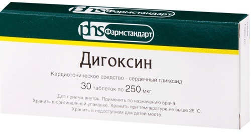
| Cardiac glycosides | Digoxin, Korglikon | The drugs help eliminate arrhythmias. The medicine should be taken before meals or after meals. The tablets should be swallowed whole and washed down with a sufficient amount of liquid. The first dose should be taken in a volume of 0.5-1 mg, then every 6 hours for 0.25-0.75 mg for 2-3 days. The maintenance dosage is 0.125-0.5 mg 1-2 times a day. |
| Beta-blockers | Diltiazem, Carvedilol | Medicines reduce the contraction of the left ventricle. Adult patients are prescribed 1 tablet 1-2 times a day. The drug is swallowed whole and washed down with a little water. |
During the course of specially selected therapy, patients should also follow the strict recommendations of the attending physician:
- Completely give up bad habits (alcoholic beverages, cigarettes).
- Always dress for the weather to prevent hypothermia or overheating.
- Avoid emotional situations, nervous strain, severe stress.
- Eating a specially formulated diet to reduce the strain on a diseased heart.
- Reduce the intensity of physical activity.
Compliance with the listed recommendations will help not only achieve a positive result in treatment, but also prevent the emergence of serious complications.
Indications and types of operations
The indication for surgical intervention is the second degree of pathological changes in the area of the tricuspid heart valve. The same applies to the lack of positive dynamics after drug therapy.
The tricuspid valve of the heart, depending on the location of pathological foci, is operated with the following methods:
| Name | Description |
| Annuloplasty | During medical procedures, the fibrous ring of the tricuspid valve is mobilized. U-shaped seams are applied to the extended commissures. |
| Semicircular annuloplasty | The operation is performed in case of strong expansion of the fibrous ring of the tricuspid valve. The surgeon uses a purse string suture. |
| Bicuspidization | Suture annuloplasty of the tricuspid valve, which involves the imposition of U-shaped sutures on the fibrous ring in the posterior cusp region. Teflon gaskets are used for this operation. |
| Prosthetics | A surgical treatment that is more commonly used for tricuspid stenosis or insufficiency. During the operation, the surgeon excises all the main valves and installs artificial prostheses in their place. |
Surgical treatment is carried out using an artificial apparatus that maintains blood circulation in the patient's body. The method of operation is selected taking into account the anatomical characteristics of the patient's body and the provoking factors that caused the development of the pathological mechanism.
Prognosis for pathologies
In most cases, progressive pathological processes with damage to the tricuspid valve lead to serious complications within 5-10 years.
Among the dangerous consequences, it is necessary to highlight:
- thromboembolism of the pulmonary vessels;
- fatal atrial fibrillation;
- congestive heart failure;
- the occurrence of ascites;
- damage to the liver or blood vessels of internal organs;
- the development of severe pneumonia;
- the occurrence of gastrointestinal bleeding due to portal hypertension.
As a result of a secondary infectious lesion of the human body against the background of defects, the risk of complete cardiac arrest increases. Life expectancy for patients with tricuspid valve disease is 20-30 years. It is important to be constantly under the supervision of a physician.
Timely diagnosis of the location of the pathological focus and treatment of the tricuspid heart valve is the main prevention against complications and consequences.
At an early stage of dysfunctions with disorders, drug therapy will help to cope. Late stages of the disease require surgery. Modern medicine offers enough methods to avoid death and prolong a person's life.
Valve Disease Videos
Why are heart valve defects dangerous?


