
Human skin - this is the largest organ, the weight of which can reach 10 kg, and the total area - up to 2 m2. Its derivatives create the necessary conditions for the normal functioning of internal organs. The histology of the skin, or the interchange of microelements between different tissue layers, ensures the normal vitality of the whole organism.
Record content:
-
1 Layers and composition of the skin
-
1.1 Epidermis
- 1.1.1 Basal layer
- 1.1.2 Prickly layer
- 1.1.3 Granular layer
- 1.1.4 Shiny layer
- 1.1.5 Stratum corneum
-
1.2 Dermis
- 1.2.1 Papillary layer
- 1.2.2 Mesh layer
-
1.1 Epidermis
-
2 Cellular composition of the epidermis
- 2.1 Keratinocytes
- 2.2 Melanocytes
- 2.3 Intraepithelial macrophages
- 2.4 Merkel's tactile cells
-
3 Derivatives of leather
- 3.1 Sweat glands
- 3.2 Sebaceous glands
- 3.3 Merocrine glands
- 3.4 Apocrine glands
- 3.5 Sebocytes
- 3.6 Hair
- 3.7 Nails
- 4 Skin types
- 5 Blood supply to the skin
-
6 Function of the skin
- 6.1 Protective
- 6.2 Thermoregulatory
- 6.3 Excretory
- 6.4 Regulation of water-salt metabolism
- 6.5 Blood deposition
- 6.6 Endocrine
- 6.7 Receptor
- 6.8 Immunological
- 7 Skin regeneration
- 8 Skin Videos
Layers and composition of the skin
The skin consists of 3 layers:
- dermis;
- epidermis;
- hypodermis.
Also between the dermis and the epidermis there is a dermo-epidermal junction, in which the processes of oxygen exchange, supply of nutrients and purification at the cellular level take place.
Epidermis
The skin and its derivatives (histology studies the importance of the skin in the general life support system) perform a barrier function in protecting the body. The epidermis, having different thickness (from 0.1 mm on the eyelids to 3 mm on the soles), makes up 17% of body weight and consists of 5 layers, in which the most important skin regeneration process takes place - renewal cells.
Basal layer
The basal layer consists of 1 cell row and is bordered by the dermis. This is where cell division begins, where they rise to the upper layers of the skin.
Passing the entire path of differentiation, they turn into keratin, which in the stratum corneum protects the skin from external factors. In the event of any damage to the skin, the process of cell division occurs faster, due to which the regeneration of damaged tissues occurs.
Prickly layer
The next layer of skin is prickly. He is responsible for starting the synthesis of keratin. Together with the basal layer, uniting in the germ layer, they create conditions for the primary activation of cells, during which they acquire the form of "thorns". 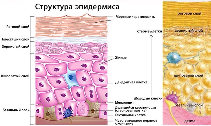 This layer has 4 to 7 cell rows, therefore it is considered the largest part of the epidermis.
Granular layer
The granular layer is characterized by the fact that keratinization of the epithelium is read in it - the synthesis of keratolinin and filaggrin cells, after which they turn into keratin. Depending on the part of the body, this layer consists of 1 to 4 rows of cells.
The main function of the granular layer is the release of fats, which in the stratum corneum hold the keratin cells together, providing along with them protection from the external environment and dehydration.
Shiny layer
The shiny layer of the epidermis is present only on the thickest areas of the skin - palms and soles - parts of the body with a pinkish tint. Lacking clear contours, the lustrous layer contains from 2 to 4 rows of cells that are involved in skin renewal.
Stratum corneum
The stratum corneum of the epidermis is the upper part of the skin that is in contact with the external environment. Being a dense barrier of scales, this layer performs a protective function against the ingress of foreign microorganisms and other destructive factors.
The main element of this layer - horny scales - are dead keratin - the final form of cells in the process of epidermal regeneration.
Dermis
The second and thickest skin layer is the dermis. In its layers (papillary and reticular), various components of the skin are formed, which are responsible for its elasticity and durability.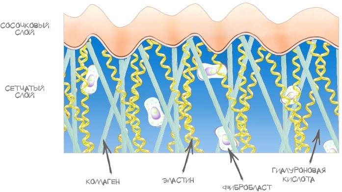
The dermis provides the epidermis with essential nutrients and oxygen, and its main components - collagen and elastin - are responsible for maintaining the elasticity of the skin, while fibroblasts, macrophages and lymphocytes, occupy an important place in the work of immunological function skin.
Papillary layer
The papillary layer of the dermis has an oblong shape in the form of papillae, which penetrate into the layers of the epidermis, due to which there is no clear distinction between different layers of the skin.
The connective tissue of the papillary layer consists of thin collagen fibers and cells:
- fibroblasts;
- fibrocytes;
- macrophages;
- lymphocytes.
Depending on the part of the body, the papillary layer can have a different thickness. Basically, the cells of the papillary layer are involved in the implementation of the immunological function of the skin, helping to produce antibodies.
Mesh layer
The reticular layer of the dermis is the main and densest part of the skin, which performs a supporting function for all the layers formed above. In structure, the mesh layer is a fibrous tissue of powerful collagen bundles and a network of elastic fibers that provides skin strength.
Cellular composition of the epidermis
Skin and its derivatives (histology studies the patterns of cell division in different skin layers) are the result of a complex process of cell formation in the layers of the epidermis.
The most important cells involved in the synthesis of the epidermis are:
- keratinocytes;
- melanocytes;
- macrophages;
- lymphocytes.
Keratinocytes
Keratinocytes are the main elements of the epidermis that contain the protein keratin. Together with other proteins (collagen and elastin), keratin forms a protective layer of the skin, giving it strength and elasticity.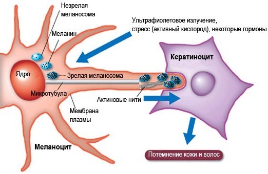
The production of keratin by the epidermis is a natural process of the body's defense against external factors.
With normal cell metabolism, the cyclical dying off of old cells occurs for 28 days - 3 months (depending on the location and age of the skin). In the case of excessive production of keratinocytes by the body, keratinized layers of tissue appear on the skin, and calluses and growths are also formed.
Keratinocytes make up 85% of the epidermal cells in all its layers and perform functions such as the formation of a protective barrier and the synthesis of vitamin D.
The process of keratinization is the cyclical production of keratin and fats by the epidermis in the upper stratum corneum, which ultimately gives strength to the surface layer of the skin.
Melanocytes
Melanocytes are protein cells of the basal layer of the epidermis, making up up to 10% of the entire layer, where the amount of this protein depends on the color of the skin. Melanocytes are involved in the production of melanin, which protects the skin from ultraviolet radiation.
Intraepithelial macrophages
Intraepithelial macrophages are found in the basal and prickly layers, promote the natural production of keratinocytes and participate in the body's immunological function by producing antibodies.
Merkel's tactile cells
Merkel's tactile cells are located in the papillary dermis:
- contribute to the healing processes of injuries;
- participate in skin regeneration;
- affect the tone of blood vessels.
Derivatives of leather
Skin and its derivatives (histology studies the structure and importance of skin tissues in the general system of the body's existence) perform the functions necessary for life support:
- protection from damage;
- removal of toxins;
- renewal of skin cells.
Sweat glands
Sweat glands are skin secretory cells that take part in thermoregulation and cleansing of the body through the secretion of sweat.
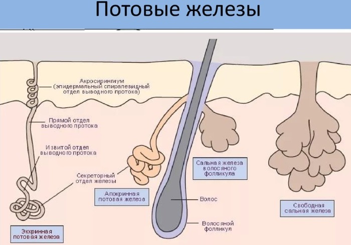
On the body of each person there are up to 4 million sweat glands, most of which are located on:
- forehead;
- face;
- in the armpits;
- palms;
- soles.
Sebaceous glands
The sebaceous glands are a derived organ of the skin that takes part in the protective function of the body. Due to the secretion of sebum, hair and epidermis are protected from external factors, as well as giving the skin softness, elasticity and durability.
The activation of the sebaceous glands is directly related to puberty and hormonal balance of the body (testosterone in men, and progesterone in women). During normal operation of the sebaceous glands, the daily rate of sebum production is 20 g.
Merocrine glands
Merocrine (eccrine) glands promote fluid secretion without disturbing skin cells. This includes the work of the salivary glands. They take part in intracellular regeneration. Their main function is thermoregulation.
Apocrine glands
Unlike the merocrine glands, where only skin secretion is secreted, during the work of the apocrine glands, dead cells are rejected in the channels of the mammary and sweat glands. These glands are located in the armpits, on the forehead, in the groin, and take part in the regeneration of the skin. They develop mainly during hormonal and puberty.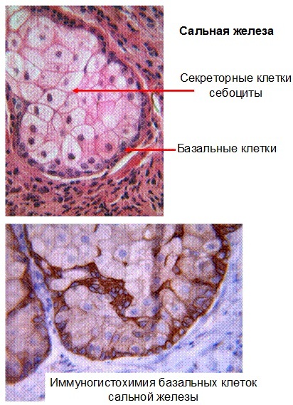
Sebocytes
Sebocytes are active secretion cells of the sweat glands, which accumulate before leaving the basal layer droplets of fat (lipids) and turn into sebum over time, removing secretions from the body through the sebaceous glands.
Hair
Hair is a filamentous protein, the growth of which occurs due to the division of tissue cells - keratin, and their color is the result of melanin production by the dermis.
Hair performs an auxiliary function in thermal insulation and protection of the body from external factors.
All the hairs that are visible on the body are already formed dead tissue that cannot be treated, and their living part is in the dermis, under the layers of the epidermis is the bulb.
It is at this level that new hair grows directly and is nourished, while the hair shaft itself no longer has a source of nourishment. With normal hair growth, the cycle of replacing an old hairline with a new one takes about 4 years.
Nails
Another derived appendage of the stratum corneum of the epidermis are nails, which serve as a protective barrier of the skin from various contacts. The nail itself does not have nerve endings and blood vessels, so a person does not feel minor mechanical damage (scratches or breaks).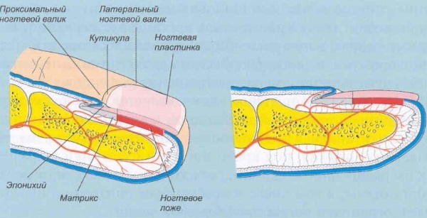
The quality of their growth is influenced by many different factors:
- balanced hormonal background;
- the presence of the required amount of minerals and fats in the body;
- normal blood circulation and metabolism.
Skin types
Skin and its derivatives (histology examines changes in the functionality of skin types in the body human), regardless of genetic predisposition and age, are characterized by different features. So, for example, the cover is divided into thin and thick.
The thickest human skin is found on the palms and soles. In these areas of the body, all layers of the epidermis have more cell rows and sometimes a slightly modified shape compared to the epidermis on other parts of the body.
Thick skin also does not have sebaceous glands, but the number of sweat glands on it is many times greater (due to which palms sweat) and more nerve endings, which is why a person has well-developed tactile Feel.
Thin skin has fewer epidermal cell rows, the shiny layer may be completely absent, the granular layer may be poorly formed, and the prickly layer is thinned to 1-2 layers of cells. Thin skin also lacks a clear delineation between epidermis and dermis. Unlike the thick one, the thin one has good hair and a developed system of sebaceous glands.
Blood supply to the skin
The skin is nourished by 3 groups of different arteries:
- cutaneous branches of arteries;
- musculocutaneous arteries;
- supra-cutaneous arteries;
These arteries are located under the hypodermis, and when they enter the dermis, they form a wide microvascular network of arteries, which carries blood to different layers of the skin.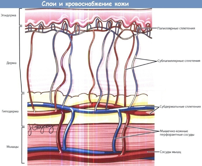
The blood supply to the skin performs a wide range of functions:
- shipping;
- adaptive;
- exchange;
- deposition (blood preservation);
- thermoregulation;
- nutrition of hair follicles, glomeruli of sweat glands and skin papillae.
Function of the skin
Skin and its derivatives (histology defines it as a complex organ of the human body) play the role of protecting internal organs from the external environment, and also many other auxiliary functions of the synthesis of useful substances, such as cleansing the body of harmful substances and preserving useful elements.
Protective
The protective function of the skin is the main and most extensive, its purpose is aimed at protecting the body a person from mechanical, chemical and thermal damage during interaction with the environment environment.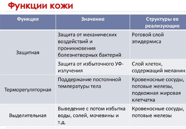
Due to the fact that the skin is practically impenetrable, it also serves as the main protection of organs from the penetration of pathogenic microorganisms inside, causing diseases and any deterioration of immunity.
Thermoregulatory
The thermoregulatory function of the skin ensures the preservation of the required temperature state of the body for its full existence.
This feature works bi-directionally:
- provides the required internal body temperature (36.6);
- protects against higher or lower ambient temperatures.
The thermoregulatory function of the skin affects the increase or decrease in the volume of internal organs, controls the state of blood vessels, sweat glands and subcutaneous adipose tissue. Also, the skin emits 80% of the heat that occurs during the work of internal organs (muscle contraction and oxidation of substances), thereby regulating the microclimate of the human body
Excretory
The excretory function of the skin, together with the work of the kidneys, removes various substances from the body.
So, for example, the skin glands, together with sweat, remove salts and other elements (urea, uric acid, ammonia, salt, toxic trace elements, drugs, as well as ions of potassium, calcium, sodium and chlorine), which enter the skin layers with blood and lymphatic system. Moreover, if the condition of the kidneys is disturbed, the excretory function is completely taken over by the skin.
Regulation of water-salt metabolism
Regulation of water-salt metabolism is a side effect of thermoregulation of the body's state, which the skin performs together with the kidneys and lungs. The excretory function removes excess water and salts from the body, thereby providing the necessary acid-base environment for the whole body to work, as well as a balanced concentration of nutrients in it and microelements.
Blood deposition
Another important function of the skin is to create a reserve of blood, or deposit, which is not involved in the general circulation throughout the body. Since human vessels are not able to retain and store blood, for emergency recovery normal circulation of blood supply, this function is taken over by some organs - skin, spleen, liver and lungs.
Thus, the erythrocyte depot retains up to 1 liter of blood in the vessels and upper layers of the skin, which, when are pumped into the circulatory system to quickly restore normal blood circulation the body.
Endocrine
The skin is an important part of the endocrine system of the body, and synthesis and production of many microelements: hormones, vitamins, which are carried throughout the body with blood, lymph and intercellular liquid.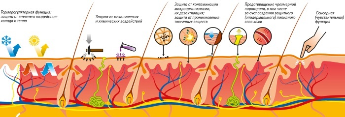
The skin absorbs and synthesizes many useful substances, as well as the direct production vitamin D - a function characteristic only of the endocrine function of the skin under the influence ultraviolet radiation.
As a result of the endocrine activity of the skin, some hormones arise: for example, testosterone is synthesized into dihydrotestosterone, and androgens into estrogens. As the body ages, the skin remains the only source that synthesizes sex hormones.
It contains endocrine functions associated with the metabolism of hormones and vitamins by the body:
| Hormones, vitamins | Function |
| Androgens | Regulation of many processes in the skin layers, for example: the formation of the work of the sebaceous glands, the effect on the hair growth cycle and the processes of skin repair. |
| Estrogens | A hormone that is formed directly in skin cells, it also regulates aging and skin pigmentation, hair growth and sebum production. |
| Thyroid hormones | Regulates normal hair growth and skin condition. |
| Corticosteroids | Manages inflammation and dermatitis, promoting faster healing of lesions |
| Catecholamines | In the cells of the epidermis, hormones such as norepinephrine and adrenaline are synthesized, which are responsible for the normal intercellular activity of the skin. |
| Corticoliberin and proopiomelanocortin | They appear in the skin layers under the influence of ultraviolet radiation, they provide anti-stress protection of the skin, increase tissue endurance. |
| Vitamin D | It is synthesized under the influence of ultraviolet radiation, regulates the exchange of minerals, as well as their normal entry into the blood and cells. |
| Vitamin A | In combination with minerals, it protects the body from colds, skin and eye diseases. |
The work of the endocrine system performs an important function of adaptation of the body to different external and internal conditions.
Receptor
The receptor function of the skin, due to the many nerve endings throughout the body, provides the perception of signals from the outside world. This characteristic affects the sensitivity to pain and other external touch and temperature perception.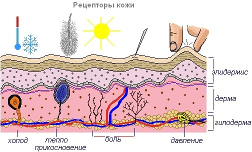
Although the receptors of the nervous system are located throughout the human skin, they are most distributed on the surface of the palms, soles, fingers, lips and genitals. The receptor function of the skin provides tactile, heat and pain sensitivity.
Immunological
The immunological function of the skin is closely related to the development of the general immunity of the body, both congenital and acquired. The main task of the skin in the formation of immune defense is participation in the production of antigens and acceleration of cell migration. Such activation of skin activity is activated in case of any violations of the integral cover, both infectious and non-infectious.
When various foreign bodies or substances (viruses, bacteria) enter the body, the skin performs an important part of a wider immunological a function aimed at capturing and transporting foreign bodies, producing antigens in order to restore normal life activity organism.
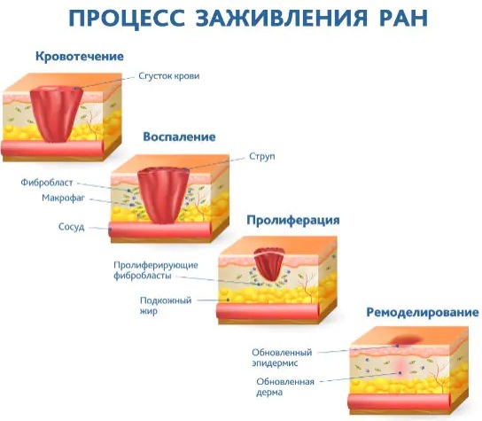
In the case of a non-infectious disorder of the skin (any injury), the injury is healed, in the process which new tissues are formed due to the migration ability of cells, which provide the regeneration process skin.
It is also important to note that the immunological function of the skin, due to age-related changes in the body, loses specific antigens, and some cells responsible for the regeneration of damage become defective. Because of this outcome, the high possibility of skin infections and the occurrence of malignant neoplasms in old age increases.
Skin regeneration
Skin regeneration is a physiological process of skin tissue renewal, which is of 2 types:
- reparative skin restoration - starts after mechanical and chemical damage to the upper layer of the epidermis;
-
physiological skin regeneration Is a cyclical process of skin cell renewal, which occurs as a result of the division of the lower cells of the epidermis and their advancement into the upper layers of the skin, where old cells give way to new ones.
Derivatives of the skin are highly dependent on the speed of the cell regeneration process. The histology of skin tissues shows that rapid tissue renewal increases the ability to completely remove traces of acne, pigmentation and wrinkles. On the other hand, poor regeneration is the result of many factors affecting the condition of the skin.
These include:
- deterioration of general immunity, hormone failure;
- long-term psychological destructive states (stress, anxiety, insomnia);
- physical exercise;
- poor nutrition.
Regardless of the general condition of the skin, the process of natural regeneration is significantly influenced by the age of a person. So, for example, the skin under 25 years old is renewed under normal circumstances in 28 days, and after 60 years - in 2-3 months.
The skin and its derivatives are one of the most important elements for the normal existence of the whole organism, the disruption in the work of which causes a long process of regeneration of the skin.
Regardless of the fact that recovery can be painless for the person himself, the histology of tissue layers is a complex process of establishing the necessary water, hormonal and cellular balance in all layers skin.
Skin Videos
Lecture. Leather and its derivatives:


