The knee has a little unstable structure. However, it is capable of supporting a person's full body weight when standing and much more when walking or running. Therefore, problems with the knee joint occur frequently in people of all ages. Understanding the anatomy of this support apparatus structure will help you understand what is happening and how knee problems develop.
Record content:
- 1 Knee joint structures
-
2 What is cartilage for?
- 2.1 Ligamentous-tendon system
- 3 Capsule part of the joint
- 4 Synovial and fibrous membrane
- 5 Muscular apparatus
- 6 Nervous system
- 7 Knee biomechanics
-
8 Diseases of the knees
- 8.1 Bursitis
- 8.2 Chondromatosis
- 8.3 Tendinitis
- 8.4 Tendinosis
- 8.5 Cyst
- 8.6 Koenig's disease
- 8.7 Schlatter's disease
- 8.8 Gonarthrosis
- 8.9 Hallux valgus and varus deformity
- 9 Diagnosing knee problems
- 10 Treatment
- 11 Knee Video
Knee joint structures
The knee consists of 3 articular compartments surrounded by a durable soft tissue sheath including:
- synovium;
- joint capsule;
- muscle;
- tendon;
- bag;
- subcutaneous tissue;
- skin.
 The complex interconnection between these structures gives the knee the function of the main weight-bearing joint of the body, which allows it to be more flexible, rotate, and have a high impact load. The joint allows the legs to bend and straighten, which is necessary for walking, going up and down stairs, running, jumping, and going from sitting to standing.
The knee joint (human anatomy is descriptively presented in the article for reference) bends around the blood vessels that pass around the knee along the tibial nerve along the back of the leg. The popliteal artery and popliteal vein are the main vessels supplying the legs and feet. The popliteal artery carries blood to the leg and foot, and the vein returns blood to the heart.
What is cartilage for?
The knee joint includes 2 main types of cartilage (articular and meniscus):
-
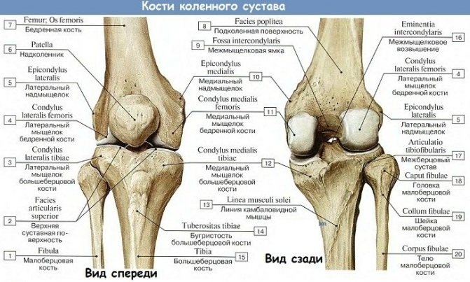
Articular cartilage covers the bony surfaces facing the cavity, and therefore is inextricably linked with them. It is durable and non-vascular, ensuring smooth articulation of 2 bones. In the knee, it is located at the proximal end of the tibia, the distal end of the femur, and at the back of the patella. Friction of the patellar cartilage against the distal femur can cause a small crackling sound known as crepitus. More often this happens under stress, for example, when a person stands up after a deep squat. This is usually not a sign of injury.
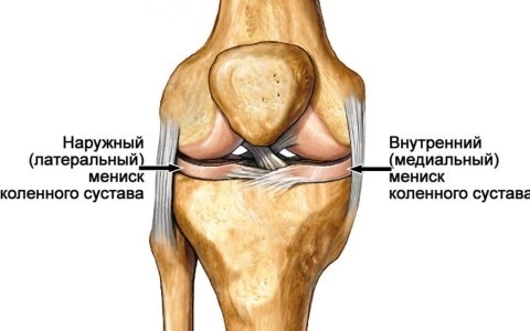
- Meniscus refers to a different type of cartilage in the knee. It is loosely attached to the tibial plateau, sits between the tibia and femur, and helps absorb shock. Each knee has 2 C-shaped menisci. The medial is located towards the midline of the body, and the lateral is towards the outside of the body. Menisci not only help absorb shock, but they also improve the coherence of the femur and tibia by distributing body weight over a larger surface area.
Ligamentous-tendon system
Ligaments are tough bands of connective tissue that connect 2 bones together. Their main function is to ensure passive stability of the joint.
The knee has several small ligaments and 4 main ones:
- cruciform anterior;
- cruciform back;
- median collateral;
- lateral collateral.
 The cruciate ligaments form an X-shape and control the movement of the lower leg back and forth with respect to the femur, respectively, and also provide some resistance to knee rotation. Collateral ligaments span the mid and lateral sides of the knee, providing stability against valgus and varus movements of the lower leg.
Capsule part of the joint
The knee joint (human anatomy in pictures clearly represents this) is surrounded by a membrane sac, called the joint capsule. It is filled with synovial fluid that lubricates and nourishes the joint.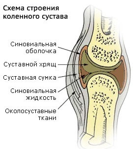
The knee joint contains about 14 of these small sacs (burs) filled with fluid. They reduce friction between the tissues of the knee and prevent inflammation.
Synovial and fibrous membrane
The cavity of the knee joint is lined with a tissue called synovial a shell that consists of 2 layers:
- intima adjacent to the knee joint itself;
- subintima, which is adjacent to the articular capsule located on the surface.
The synovium promotes the production of synovial fluid, which provides joint lubrication and diffusion of nutrients into the articular cartilage. The first layer contains 2 types of specialized cells, or synoviocytes, which are responsible for the production and maintenance of synovial fluid. The second layer is rich in blood, lymphatic vessels, and nerve endings.
Muscular apparatus
The knee has many muscles that affect the joint, but the main muscles that allow the knee to perform its main functions are:
- Quadriceps is a group of 4 muscles located on the front of the thigh. These are the vastus lateralis, the vastus medialis, the vastus intermediate, and the rectus femoris. They are responsible for straightening the knee.
- Hamstrings Is a group of 3 muscles that are located on the back of the thigh and allow the knee to bend. This is the biceps femoris, semitendinosus and semimembranosus muscles. The hamstrings work together to bend the leg at the knee, they are responsible for raising the limb when walking.
- Calf muscles - This is a group of 2 muscles that are located on the back of the lower leg. They work in tandem with the hamstrings to cause the knee to bend. In the calf region of the lower leg, the calf muscle extends from the distal end of the thigh through the calcaneus (Achilles) tendon to the calcaneus. The calf muscle forms the posterior muscular wall of the knee and acts as the flexor of the knee and the plantar flexor of the foot.
All of these muscles also perform functions in various joints such as the hip and ankle.
Anatomically, the knee joint has other muscles that help a person to move. These include the muscles that stretch the fascia lata, the popliteal muscle, and the articularis (skeletal) muscles. The fascia lata helps stabilize the femoral and tibial muscles.
Flexion of the knee is achieved by contraction of the popliteal muscle. A tiny muscle from the genus articularis lifts the patellar sac and the capsule of the knee joint to prevent pinching of this soft tissue during knee extension.
Nervous system
The main nerves of the knee are located at the back of the knee. This is the tibial and common peroneal nerve. They extend to the lower leg and foot, providing muscle sensitivity and control.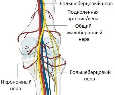
They form from a large sciatic nerve that splits just above the knee. The tibial nerve runs down the back of the leg, while the common peroneal nerve runs along the outside of the knee and along the front of the leg to the foot.
Knee biomechanics
The knee joint (human anatomy clearly shows the principle of joint mechanics) rotates according to the “helical point” principle, which allows the knees to be fully extended and flexed. Ligaments and menisci provide static stability to the muscle, while tendons provide dynamic stability.
Osteokinematics and range of motion:
- The main movement in the knee is flexion - extension. In this respect, the knee acts as a hinge joint whereby the articular surfaces of the thigh roll and slide over the surface of the tibia. During flexion and extension, the shin and patella act as one structure in relation to the femur. The 4-head muscle group is made up of four separate muscles that join together to form a single tendon. It is included in the anterior tuberosity of the tibia. A 4-head contraction pulls the patella up and extends the knee. Range of motion: extension 0˚. The hamstring muscle group located on the back of the thigh flexes the knee and also provides stability on either side of the joint line. Range of motion: 140˚ flexion.
The internal secondary movement is the rotation of the lower leg in relation to the femur, but it is possible only with the knee bent.
Arthrokinematics of the knee joint is based on the rules of concavity and convexity, when the articular the surface of the thigh is convex, and the tibial is concave and is described in terms of open and closed chains:
- Open kinetic chain. With knee extension, the tibia slides forward over the thigh. More specifically, from 20˚ of knee flexion to full extension, the tibia rotates outward. During knee flexion, the tibia slides posteriorly over the femur, and from full knee extension to 20˚ flexion, the tibia rotates inward.
- Closed kinetic chain. During knee extension, the femur slides posteriorly over the tibia, from 20˚ of knee flexion to full extension, and the femur rotates internally on the stable tibia. During knee flexion, the femur slides anteriorly on the tibia and from full knee extension to 20˚ flexion, while the femur rotates outwardly on the stable tibia.
The 'screw-down' mechanism is considered a key element of knee stability, which is the rotation between the tibia and the femur.
Diseases of the knees
The knee joint is exposed to great stress, and hence the multiplicity of its diseases and disorders.
- Inflammation of soft tissues - bursitis, synovitis, tendonitis.
- Injuries - bone fractures, rupture of ligaments, muscles, damage to cartilage, menisci.
- Knee diseases - arthritis, infections, cancer.
- Neuromuscular diseases - myalgia, myopathy, spasm, obturator nerve syndrome, femoral nerve neuropathy.
Bursitis
Knee bursitis is inflammation of a small sac filled with fluid. Bursa reduce friction and soften pressure points between bones, tendons, muscles near joints.
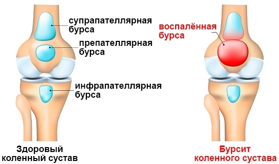
Any bursa of the knee can become inflamed, but most often knee bursitis occurs on the patella or on the inside of the knee below the joint. The inflammation occurs as a result of work that requires frequent kneeling on hard surfaces, so symptoms usually begin gradually and may worsen over time.
The symptoms of knee bursitis differ depending on which bursa is affected and what is causing the inflammation.
The main signs are:
- pain, swelling;
- decreased knee mobility or stiffness;
- red, warm, skin over the knee.
Chondromatosis
Synovial chondromatosis is a type of benign tumor. The knee is most commonly affected, but any joint can be affected. Tumors begin as small cartilaginous nodules that may appear and separate within the joint. Some tumors may be no larger than a grain of rice. Synovial chondromatosis most often occurs in adults between the ages of 20 and 50.
Symptoms may include:
- pain;
- edema;
- reduced range of motion;
- joint blockage.
The exact underlying cause of the disease is unknown.
Tendinitis
Patellar tendonitis is an injury to the tendon that connects the patella (patella) to the tibia. Patellar tendonitis, also known as "jumper's knee", is more common in athletes who play basketball and volleyball.
Pain is the first symptom of patellar tendinitis. It occurs between the patella and where the tendon attaches to the tibia.
The knee joint (the human anatomy in the pictures is presented for reference) can hurt with tendinitis at the beginning of physical activity or immediately after an intense workout. Over time, the pain gets worse and interferes with daily movements, such as climbing stairs or getting out of a chair.
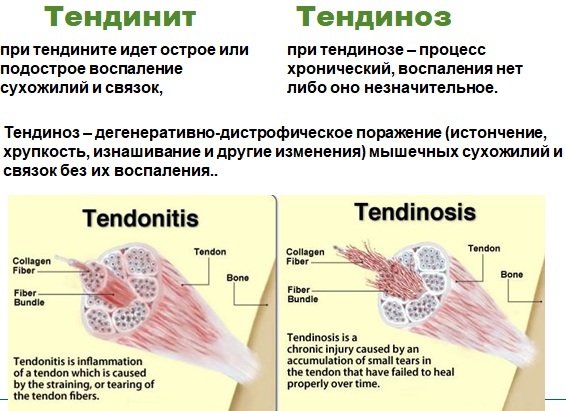
Knee tendonitis symptoms include:
- pain above or below the kneecap;
- swelling;
- pain that repeats with certain activities and is relieved with rest;
- in severe cases, the pain becomes constant (despite the rest of the joint) and can even disrupt sleep.
Tendinosis
Tendinosis is a condition characterized by inflammation of the patellar tendon that connects the patella to the tibia. Tendinosis weakens the tendon and, if left untreated, can rupture.
Symptoms:
- pain and swelling around the tendon;
- pain when jumping, running or walking, when bending or extending the leg;
- soreness behind the lower part of the kneecap.
Cyst
A Baker's cyst is a fluid-filled bladder that causes a feeling of tightness behind the knee. The pain may increase with full flexion or extension of the knee.
A Baker's cyst, also called a popliteal cyst, usually results from a problem with the knee joint, such as arthritis or ruptured cartilage. In some cases, the cyst is not painful and may not be noticed.
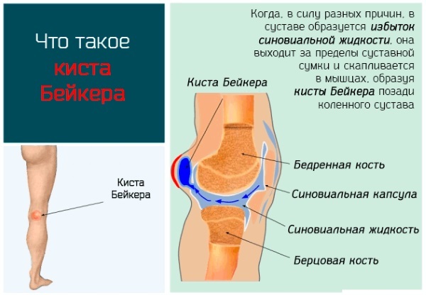
But symptoms may appear:
- swelling behind the knee and sometimes in the leg;
- knee pain;
- stiffness and inability to fully bend the knee.
Symptoms may worsen after physical activity or long standing.
Koenig's disease
Osteochondrosis of the inner condyle of the femur is a condition in which there is a limited area of necrosis of the articular cartilage and adjacent bone in the inner condyle of the femur.
The cause of this disease is not fully understood. It is believed that necrosis causes poor circulation in the branches of the arteries of the middle knee. Another point of view associates this disease with chronic knee injury. Mostly young people 20-25 years old are ill. Initially, the patient has mild aching pain in the knee joint. The pain increases with and after exercise. When palpating, pain is felt on the inner surface of the knee joint.
Schlatter's disease
Osgood-Schlatter disease is a condition that causes pain and swelling below the knee joint, where the tendon the patella attaches to the top of the tibia at a point called the tibial tuberosity bones. There may also be inflammation of the patellar tendon that extends over the patella.
The disease is caused by irritation of the bone growth plate. The bones do not grow in the middle, but at the ends near the joint, in an area called the growth plate. As the child grows, these growth zones are made up of cartilage, not bone.
Cartilage is never as strong as bone, so high levels of stress can cause the growth plate to ache and swell. The disease is most common in young athletes who play sports that require a lot of jumping or running.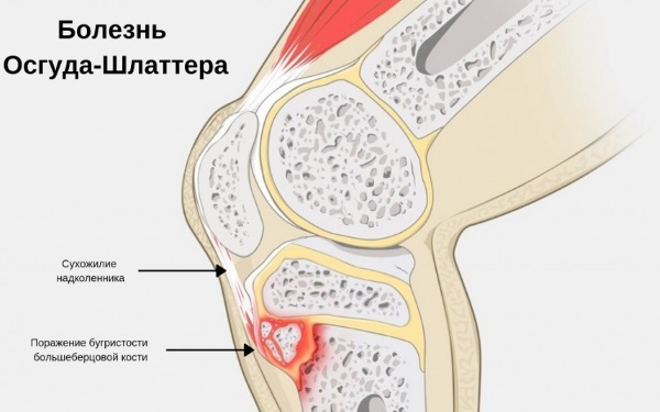
Common symptoms:
- leg or knee pain;
- swelling, soreness, or increased warmth under the knee and over the tibia;
- pain that gets worse from exercise or exertion;
- lameness after exercise.
Gonarthrosis
The knee provides the smoothest possible movement of the hips and calves. Daily stress affects the wear and tear of the knee joint. Experts call the resulting disorder osteoarthritis of the knee joint or gonarthrosis.
In the early stages, knee pain occurs only with excessive exertion. In most cases, patients complain of pain during long walks or climbing stairs. Swelling in the knee joint is often present. All these symptoms are clear signs of knee osteoarthritis (gonarthrosis).
The wear of the knee joint can be caused by a predisposition, or it can be the result of an illness or accident. The elderly are particularly affected.
Hallux valgus and varus deformity
Knee osteoarthritis can cause a valgus or varus position. In a fully aligned, correct knee joint, the mass is evenly distributed over the knee joint. But not all knees are positioned correctly. If the knee joint flexes noticeably or inward, this unevenly distributes mass throughout the joint, which is called knee displacement.
Hallux valgus is the term for the outward bending of a distal bone segment or joint. The opposite deformity - medial deviation of the distal part of the bone - is called varus. The terms varus and valgus always refer to the direction in which the distal segment of the joint points.
The first symptom is pain in the knee, leg. Sometimes it is foot or ankle pain, but more often it is knee pain.
Visual symptoms:
- slight lameness in the affected part;
- change in the volume of the thighs and legs;
- deviation of the distal end of the knee inward, which gives the impression of a bent leg.
Diagnosing knee problems
There are 3 approaches to diagnosing knee pain:
- Location of pain: Where the knee hurts, such as the front, side, or back.
- How the pain appeared: Mechanism of injury, such as sudden twisting or gradual onset.
- Specific symptoms of the patient.
In some cases, visual tests are performed:
-
X-ray. It can help detect bone fractures and degenerative joint disease.
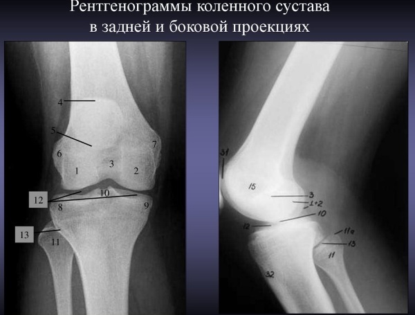
X-ray of the knee joint in two projections - CT scanners diagnose bone problems, small fractures can accurately identify gout, even if the joint is not inflamed.
- Ultrasound. This technology examines the soft tissue structures in and around the knee.
- MRI useful for detecting damage to soft tissues such as ligaments, tendons, cartilage, and muscles.
If a doctor suspects an infection or inflammation, blood tests and sometimes a procedure called arthrocentesis, in which a small amount of fluid is taken from the knee joint with a needle and sent to a laboratory for analysis.
Treatment
The knee joint (the human anatomy in this area has been thoroughly studied by doctors) can be subject to external and internal disorders, and therefore pains can be of a different nature. Treatments for knees are as varied as the conditions that cause pain. Therapy is aimed at reducing pain, restoring joint stability, relieving swelling and weakness.
| Treatment method | The ways |
| Strengthening muscles | In 99.9% of cases, muscle weakness is the main cause of knee problems, so strengthening leg muscles is one of the best all-around treatment options for knee pain. |
| Stretching the muscles | Tight muscles are a common cause of knee pain. Stretching as part of your treatment plan can help reduce joint stress. |
| Medicines | Drugs like Tylenol, Paracetamol can help reduce pain, and NSAIDs like Ibuprofen can help reduce swelling. |
| Ice | Cold plays a very important role in treating knee pain in the early stages after injury or long-term problems. It reduces blood flow to the affected area by limiting internal bleeding, which reduces swelling and pain. |
| Warmly | A natural remedy for knee pain for longer-term knee problems such as arthritis, rather than after initial injuries. Heat works in 2 ways: it increases blood flow to the affected area, reduces pain and helps muscles relax. |
| Orthosis |
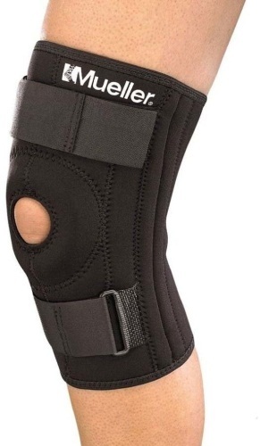 Special devices for the knee joint help to reduce the load on it, provide support and stability, especially after injuries, and reduce pain. Special devices for the knee joint help to reduce the load on it, provide support and stability, especially after injuries, and reduce pain. |
| Injections | Intra-articular injections help with long-term problems such as ruptured cartilage and arthritis. 2 types of injections are used: corticosteroids to relieve pain and swelling, and Synvisc injections, prosthesis synovial fluid to improve lubrication of the joints, Recent injections are especially helpful in treatment arthritis. |
| Acupuncture | Helps reduce knee pain. |
| Knee surgery | If other treatments for knee pain have not worked, knee surgery may be needed. This can be a minor surgery such as arthroscopy or a complete joint replacement. |
Some treatments work well in the early stages after a knee injury, while others work well for a long period. For therapy to be effective, treatment must be directed not only at the symptoms of the problem, but also at what causes it, so that the problem does not return in the future.
Since the knee joint is anatomically a complex structure subject to significant biomechanical stress on a daily basis, it is a frequent site of injury. Therefore, any unnatural movement of a person's knee, such as twisting, twisting, sudden change in direction, or violent impact, can damage these structures.
Knee Video
Knee Anatomy: