
The heart is the main engine of the body. Throughout a person's life, it constantly beats and reaches maximum performance under increased loads. The main thing that is needed for this is oxygen, which is delivered according to a certain scheme through the coronary arteries.
Record content:
- 1 Characteristic
- 2 Functions and properties
-
3 Anatomy and structure
- 3.1 Left and right arteries
- 3.2 Left-sided coronary artery
- 3.3 Right coronary artery
- 3.4 Artery structure
- 3.5 Collateral circulation
-
4 Possible diseases and pathologies
- 4.1 Heart attack or myocardial infarction
- 4.2 Angina pectoris
- 5 Video about the arteries of the heart
Characteristic
The heart is supplied with blood arteries of the coronary blood system, which branch off from the aorta, run along the outer surface of the heart, forming a plexus in the form of a crown. The main tasks of the coronary arteries are the delivery of blood to the region of the right and left ventricles of the heart.
Arteries diverge into capillaries, due to which the myocardium (heart muscle) is supplied with oxygen, and then converge again into coronary veins to divert deoxygenated blood back to the right atrium, where the blood will be re-oxygenated through the pulmonary circuit.
Functions and properties
The coronary arteries of the heart (a diagram of the structure of the heart complex is available on the site) are isolated on their way small branches, due to which there is a supply of oxygen and nutrients to the cells of the heart muscles.
With the help of oxygen and energy, the heart performs all of its tasks, including the delivery of electrical impulses and conduction, as well as pumping and supporting vital circulation.
The coronary arteries provide the delivery of oxygenated blood to the heart muscle. There is high pressure in the area of coronary blood flow. During systole, up to 15% of blood flows to the heart muscle, and during diastole - about 80%. Coronary blood flow is 250 ml / min, and oxygen consumption is 9.5 ml per 100 g.
The main task of the right coronary artery is to ensure proper blood circulation in the myocardium.
The left coronary artery and its branches play a critical role in ensuring that the muscles of the heart itself are supplied with oxygen. In particular, it provides most of the lower chambers of the heart, as well as the left atrium and atrial appendage, pulmonary artery, and aortic root.
Anatomy and structure
The heart is a pump made up primarily of heart muscle cells that are incredibly active throughout life. Like all other cells, the cardiomyocyte requires a reliable supply of oxygen and nutrients, and also a way to remove slags, so it needs a specialized, complex and extensive coronary circulation.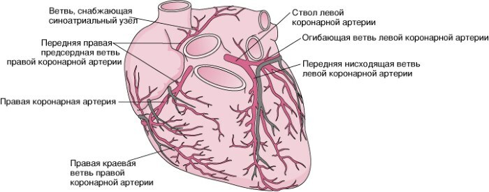
And because of the critical and almost continuous activity of the heart throughout life, this need for blood supply is even greater than for a typical cell.
The heart does not receive from the blood that is in its chambers, nutrition and oxygen, because this is hindered the thickness of the middle heart layer, myocardium, and the waterproof shell of the inner layer of the heart wall, endocardium.
For this, it acquired its own vessels, which make up the coronary arterial system. It includes 2 main arteries: left and right. The first, soon after its inception, is divided into 2 main branches. Thus, one usually speaks of 3 main functional coronary arteries belonging to the heart.
Left and right arteries
The cardiac circulatory system receives blood through the left and right arteries. They originate to the right and left of the aortic sinus (Valsalva sinuses).
Sinuses are protrusions of the walls at the beginning of the ascending aorta, corresponding to the semilunar flaps of the aortic valves. In the right sinus of the aorta is the opening of the right coronary artery, and the opening of the left is in the sinus of the left aorta.
Left-sided coronary artery
The left-sided coronary artery supplies blood to the left and posterior sides of the heart. It travels a short path through the anterior part of the heart, dividing into the left anterior descending artery and the left circumflex artery. It is about 4.5 mm in diameter and only 1-2 cm long to the point of division.
Moving between the pulmonary trunk (connecting the heart to the lungs) and the appendage of the left atrium, it begins to split into 2 final branches:
- the circumflex branch, which wraps around the heart, provides blood flow to the muscles on the back of this organ;
- left anterior descending branch, as an extension of the left coronary artery. The branch, going down, supplies blood to the left ventricle and the muscles of the anterior part of the heart.
Coronary arteries of the heart
In about 0.5% of people, the left coronary artery is missing. In this case, the circumflex and left anterior descending artery extends directly from the aorta.
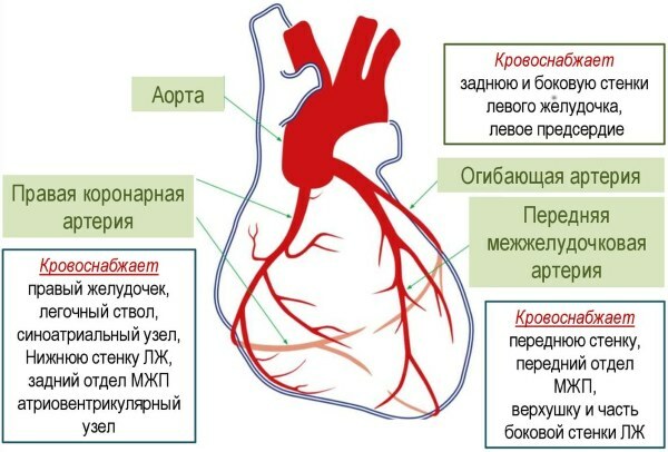
Basic facts about the left coronary artery | |
| Origin | Left-sided aortic sinus of the ascending aorta. |
| Main branches | Circumflex artery Left anterior descending artery |
| Where does the blood deliver | Left atrium, part of the right ventricle, left ventricle, anterior 2/3 of the interventricular septum, atrioventricular bundle, sinoatrial node (40%). |
Right coronary artery
The right coronary artery is the main artery supplying the heart and its branches are the main sources of blood for the right ventricle and atrium of the heart.
The artery travels downward through the right atrioventricular groove separating the right atrium and right ventricle before curving backward. It divides into 2 main branches: the right marginal artery and the posterior descending artery, which supply blood to the lower part of the heart.
Two main branches extend from the right coronary artery within 1 mm of the aortic exit:
- Cone artery. It directs blood to the ventricular outflow, a sort of gateway for blood to the main arteries of the heart.
- A branch of the sinus-atrial node. It leads to the sinoatrial nodular artery, which runs behind the right atrium and then surrounds the superior vena cava, the vessel that carries deoxygenated blood to the heart.
The right coronary artery is further divided into a right marginal branch and a posterior ascending artery. The first branch runs along the right side of the heart and supplies the right atrium and ventricle. The posterior ascending artery delivers blood to the lower part of the heart.
Subsequently, the artery branches off at the nodal point of the heart and supplies the atrioventricular node, as well as the bundle of His, both of which are associated with the coordination of electrical signals in the heart.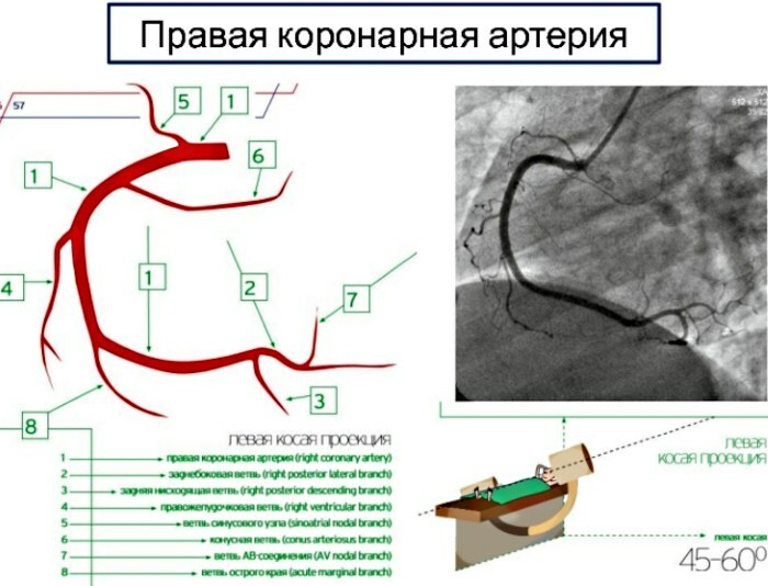
| Origin | Right aortic sinus of the ascending aorta |
| Main branches | Front branch Marginal branch Lower branch |
| Where does the blood deliver | Right atrium, right ventricle, phrenic part of the left ventricle, atrial and interventricular septum, atrioventricular node, and sometimes sinoatrial node. |
Artery structure
An artery is an elastic blood vessel, the wall of which consists of 3 layers:
- Tunica adventure - strong outer covering of the arteries. Consists of connective tissue, as well as collagen and elastic fibers. These fibers allow the vessels to stretch to prevent excessive expansion due to the pressure that the blood stream exerts on the walls.
- Tunica media - the middle layer of the walls. It is composed of smooth muscle and elastic fibers. This layer is thicker in arteries than in veins.
- Tunica intima - the inner layer. In arteries, this layer consists of an elastic membrane lining and a smooth endothelium (a special type of epithelial tissue) covered with elastic tissue.
 The coronary arteries of the heart (the diagram presented on the site allows you to see the segments of the vessels) are histologically similar to the arteries visible throughout the body. The inner shell consists of a continuous endothelium and an associated subendothelial connective tissue space, bounded from the outside by an internal elastic membrane.
Smooth muscle cells and elastic plates occupy the middle membrane, and the outer coating consists of cells and fibers of connective tissue. Coronary arteries passing through the epicardium are characterized as "elastic" arteries, although the number they have more smooth muscle cells, and the number of elastic fibers is less than in other elastic arteries.
The branches of the main epicardial arteries that penetrate the myocardial wall are classified as "muscle" arteries, which in turn give rise to arterioles and eventually the capillary bed.
The diagram of the coronary arteries does not contain anastomotic connections between the large coronary arteries in the heart, but they are between arterioles from 20 to 150 μm in the diameter of the right and left coronary arteries or between the branches of each of them, without intermediate capillaries.
These collateral joints are of medical importance in heart disease. Due to their small diameter, they cannot quickly provide a detour sufficient to bypass the blood flow in the event of sudden coronary artery disease.
If one of the coronary branches is blocked, the anastomoses cannot form a sufficient bypass circuit - a blocked coronary artery always means a heart attack.
Coronary vessels vary individually in size. In some people, for example, the left coronary artery supplies nearly the entire heart.
Collateral circulation
In case of blockage of the main blood vessel, collateral vessels begin to work. Collateral circulation is a network of tiny blood vessels. When the coronary arteries narrow to the point that blood flow to the heart muscle is restricted (coronary artery disease), collateral vessels can enlarge and become active.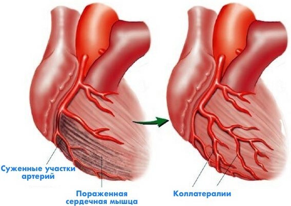
This allows blood to flow around the blocked artery to another nearby artery or to the same artery, bypassing the blockage, protecting the heart tissue from damage. The coronary circulation is not devoid of collaterals, but in healthy people they are very poorly developed, therefore they do not represent a rescue valve in case of sudden vascular obstruction.
Possible diseases and pathologies
The central role that the left coronary artery plays in the functioning of the heart means that a disease or disruption of its or its branches can lead to very serious problems. Particularly when the walls of the arteries narrow due to hardening and buildup of plaque (atherosclerosis), decreased normal blood flow can lead to coronary artery disease.
In these cases, especially if the blockage becomes complete, the muscles of the heart do not receive enough oxygen, a condition called ischemia develops. Atherosclerosis, if progressed, causes the vessels to harden, severely restricting blood flow.
This damages part of the heart and affects the amount of blood flow to the rest of the body. In extreme cases, a complete blockage here can lead to a heart attack and possibly death.
Heart attack or myocardial infarction
This is irreversible damage to the heart muscle. "Myo" means muscle, "cardiac" refers to the heart, and "heart attack" means tissue death due to lack of blood supply.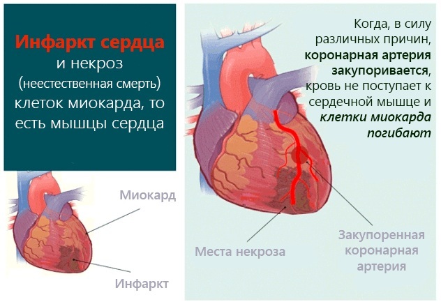
If a blood clot completely blocks the blood supply to the heart muscle, called a coronary thrombus or occlusion, the heart muscle "starves" for oxygen and nutrients (this is called ischemia) in the area below blockages. Acute coronary syndrome may occur within a short time.
This is the name given to 3 types of coronary artery disease that are associated with the sudden rupture of plaque inside a coronary artery:
- unstable angina;
- myocardial infarction without ST segment elevation;
- myocardial infarction with ST segment elevation.
Not all people who have had heart attacks have the same symptoms. Some experience mild pain, others more severe. Sometimes there may be no symptoms at all or sudden cardiac arrest. However, the more signs, the higher the likelihood of a heart attack. A heart attack is less common due to a spasm of a coronary artery.
During a coronary spasm, the arteries constrict or spasm, causing a lack of blood supply to the heart muscle (ischemia). It can occur at rest and even without significant symptoms in the coronary artery. If the spasm occurs over a long period of time, a heart attack may occur.
Some attacks start immediately, but many people have warning signs hours, days, or weeks before. The earliest warning may be recurrent chest pain or pressure (angina) caused by physical activity and diminished at rest.
Coronary arteries of the heart (a drug regimen is required during treatment) due to insufficient blood flow caused by a blood clot are prone to thrombosis. In this case, thrombolytic drugs are prescribed to dissolve the clots. Antiplatelet drugs and aspirin are commonly used to prevent blood clots from forming again.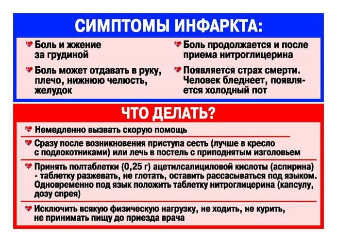
If the lack of blood flow is associated with fixed stenosing coronary artery disease due to atherosclerosis, blood flow can be improved within this vessel:
- by placing a stent inside the vessel to expand the lumen;
- when using a balloon in the process of intracoronary angioplasty to stretch the vessel;
- bypassing the diseased vessel with a vascular graft.
The coronary arteries of the heart (a schematic view can be seen in the article) due to lack of oxygen can significantly narrow.
In particular, during physical exertion, stress, the heart can no longer transport enough blood through the body, and a variety of symptoms arise:
- neck or jaw pain;
- shoulder or arm pain;
- fast heartbeat;
- shortness of breath with physical activity;
- nausea and vomiting;
- sweating
Some people have no symptoms, in such cases they speak of silent ischemia. The disease not only affects the electrical and mechanical function of the heart, but it also often causes severe and debilitating chest pain known as angina.
Angina pectoris
The disease occurs when one or more of the coronary arteries become narrowed or blocked. Discomfort with angina pectoris may be mild at first, and then gradually worsen or a sharp development of the disease occurs.
Angina pectoris usually resolves quickly. However, it could be a sign of a life-threatening heart problem. Angina can usually be controlled with medication and lifestyle changes.
Angina is usually felt as a burning or pressing pain in the chest. The main pain occurs under the sternum, and can spread up towards the throat and jaw. Discomfort is sometimes felt in the left hand and sometimes in both hands. Patients with angina pectoris often develop cold sweats. Other symptoms may include shortness of breath, dizziness, and nausea.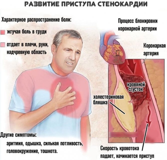
Doctors divide angina pectoris into 2 types:
- Stable angina. Chest pain occurs during physical activity or after intense emotions. Exercise in cold weather or after a heavy meal is more likely to cause angina.
- Unstable angina - the symptoms are less predictable. The pain occurs at rest, during sleep, or with minimal exertion.
If coronary heart disease is predominantly atherosclerotic in nature, then inflammatory coronary artery disease can be just as life-threatening heart disease at all ages groups. Vasculitis of the coronary arteries is a rare cause of acute myocardial infarction.
It is a general term for inflammation of the blood vessel walls, which can lead to stenosis, occlusion, aneurysm, or rupture. It can be isolated or caused by other systemic vasculitis.
Strengthening and treating coronary arteries and other blood vessels is a holistic approach to restoring heart health that takes time and effort. Therapeutic, and especially prophylactic, regimens prescribed by a doctor must be strictly observed by patients.
Video about the arteries of the heart
Anatomy of the arteries of the heart:


