Large chest - This is a thick fan-shaped muscle that participates in the movement of the scapular-thoracic joint. Its main function is to attach or bring the hand to the body.
Record content:
- 1 Characteristic
- 2 Functions and properties
- 3 Anatomy and structure
-
4 Possible diseases and pathologies
-
4.1 Myositis
- 4.1.1 Classification
- 4.1.2 Symptoms and Signs
- 4.1.3 Causes
- 4.1.4 Treatment methods
-
4.2 Tears and tears of the pectoralis major muscle
- 4.2.1 Classification
- 4.2.2 Symptoms and Signs
- 4.2.3 Causes
- 4.2.4 Treatment methods
-
4.3 Poland syndrome
- 4.3.1 Classification
- 4.3.2 Symptoms and Signs
- 4.3.3 Causes
- 4.3.4 Treatment methods
-
4.1 Myositis
- 5 Video about the pectoralis major muscle
Characteristic
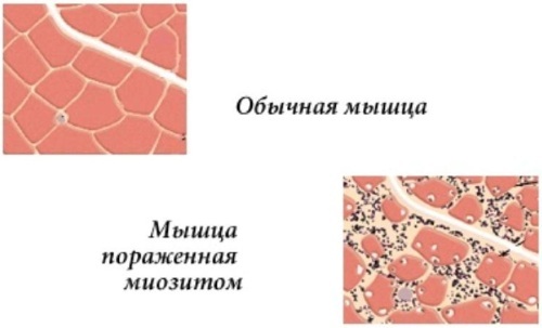
Pectoralis major is a striated skeletal muscle. They present a clear linear cross-section under the microscope that distinguishes them from smooth muscle. Skeletal muscles contract their fibers by voluntary contraction, and then relax them.
The pectoralis major muscle attaches to the humerus and lies under the pectoral fascia, which is part of the trunk fascia.
It consists of 3 parts or bundles:
- clavicular head;
- sternocostal head;
- abdominal part.
The most common muscle injuries are those involving partial rupture of a deep head or complete rupture of both heads.
Functions and properties
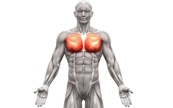
The pectoralis major muscle covers most of the chest. The widest part originates at the sternum, then is attached with the help of the heads at the top of the bony structure of the shoulder. Both heads have different functions depending on the angle of movement of the shoulder.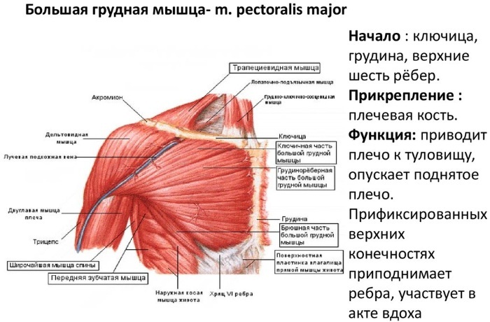
- The clavicular head helps to flex and medially rotate the shoulder joint.
- The sternocostal head originates from the sternum. This part makes up 80% of the total volume of the great muscle and provides the main part of the muscle action: adduction of the arm and internal rotation of the humerus.
Anatomy and structure
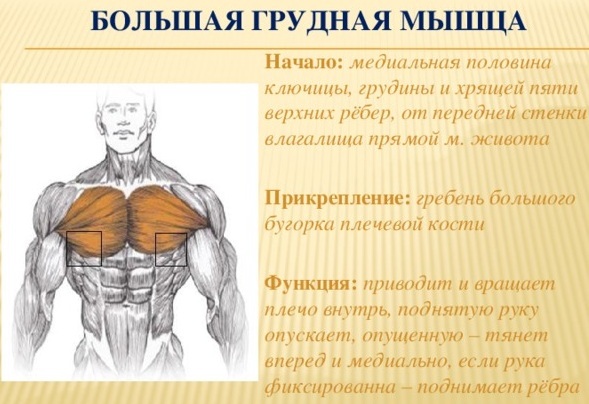
The pectoralis major is a thick fan-shaped muscle located in the upper chest that provides adduction and internal rotation of the arm.
It has 4 points of origin:
- the anterior surface of the medial half of the clavicle;
- the lateral half of the anterior surface of the sternum handle, where the 6th or 7th costal cartilage is attached;
- costal cartilage from the 2nd to the 6th rib;
- aponeurosis of the external oblique muscle of the abdomen.
From these sources, the muscle fibers converge laterally to the attachment site at the shoulder. Fibers extending from the clavicular part pass at an angle downward and to the sides, separated from the sternocostal part by a groove. Fibers from the sternocostal head pass laterally and upward. They join to form a flat tendon 5 cm high, which is inserted into the crest of the greater tubercle of the humerus.
The pectoralis muscle, together with the tendon, gives a natural appearance to the anterior axillary fold. The point serves as a reference point for surgeons during operations on the subclavian artery. The pectoralis minor muscle, which originates on the anterior surface of the 3-5th rib and attaches to the coracoid process of the scapula, lies deep in relation to the major muscle.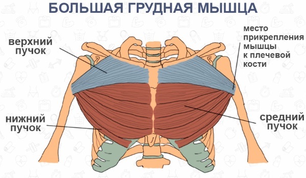
It performs the function of moving the scapula down and forward. The serratus anterior and intercostal muscles are located deep in the pectoral muscles. Above, the pectoralis muscle is bordered by the deltoid muscle. The triangular depression between the pectoralis major, deltoid muscle and the clavicle is called the interclavicular fossa (Morenheim fossa).
The pectoralis major muscle attaches to the crista tuberculi majoris (humerus). In men, it is covered with a layer of adjacent skin, subcutaneous tissue and deep superficial fascia. In women, it is covered by the chest.
Innervation occurs due to the lateral and medial pectoral nerves extending from the brachial plexus.
Blood supply is carried out by the thoracic branches of the thoracoacromial artery and the intercostal branches of the internal thoracic artery.
Both the pectoralis major and minor muscles help to move the arms forward, bring them together. The ribcage has many fibers with different angles. Each muscle acts depending on the position of the arm and fibers. This is especially true of the movement of the shoulder joint, the spherical joint between the scapula and the humerus.
For its movement, the coordinated work of various muscles is necessary. Therefore, various muscles, small and large, were laid in the structure of the human body, which are responsible for various types of movements and creating a range of motor activity of the shoulder joint, arms, shoulder blades.
Possible diseases and pathologies
Injury to the pectoralis major tendon is relatively rare. The tendon is damaged during eccentric contraction - when the external force acting on the muscle exceeds the force it can generate, and when the arm is extended and rotated outward.
The bench press is the most common cause of injury, but there are other activities that cause injury, including skiing, parachuting, football, wrestling, and hockey. Serious chest injuries usually occur in men between the ages of 20 and 40.
Symptoms are pain in the chest, shoulder, and inside the arm up to the elbow. Sometimes this pain is mistaken for heart pain.
- In women, along with chest pain, nipple soreness or hypersensitivity may occur.
- Painful sensations can spread to the ring finger and little finger, manifest when the arms are extended.
- A sensation of pain is often manifested between the shoulder blades.
- Severe pain can cause a feeling of tightness in the chest that resembles chest angina.
- Initially, the pain may be one-sided, but if left untreated, it spreads to the other side of the chest.
Muscle pain in the chest is not a specific symptom, does not indicate a specific problem, therefore, the causes should be sought in conjunction with a doctor.
Myositis
The pectoralis muscle at the point of attachment to the humerus is a flat tendon. The inflammatory process that occurs in a large muscle can affect not only muscle tissue, but also a tendon.
Injury or forceful action in athletes often leads to inflammation of the pectoral muscles and tendons.
Classification
There are several types of myositis:
- local, which affects 1 muscle group;
- polymyositis, in which several muscle groups are affected;
- dermatomyositis, an inflammatory process that affects not only muscles, but also the skin.
Symptoms and Signs
Myositis causes limitation of movement in the shoulder joint.
The main signs of myositis:
- Pain occurs as mild or moderate with physical activity.
- After 7-10 days, the inflammation intensifies and the pain becomes severe even at rest.
- The pain in the neck, shoulders, chest area intensifies.
- Muscles become tense, stiff, and responsive to touch.
- The person has difficulty getting up.
- Hoarseness or whispering appears.
- Difficulty swallowing.
Causes
The disorder is often seen in contact sports patients with ligament and muscle injuries.
Common reasons:
- Superficial trauma that caused a micro-rupture of muscle tissue.
- Lifting weights daily to increase muscle strength.
- The uncoordinated movement of the joint during muscle contraction causes excessive stress on the muscle fibers, which contributes to the appearance of microtraumas in the tendons and muscles. Such trauma follows inflammation.
- Inflammation can be due to excessive stretching of tendons and muscles during sleep or a sedentary lifestyle.
- A sharp twist of the arm can cause excessive stretching of the pectoralis muscles and tendons.
Muscle strain injuries are common in men aged 20 to 40. Rarely, but occurs in the elderly, especially after an unsuccessful fall, muscle overexertion.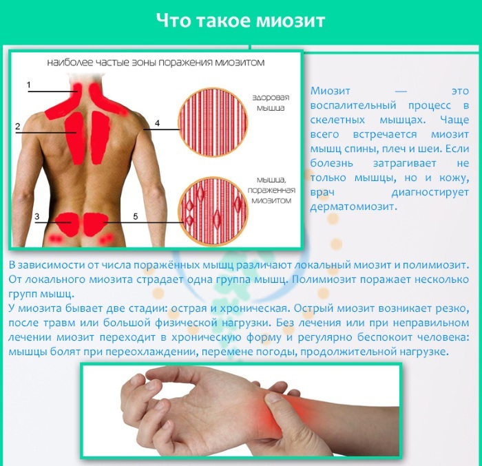
Patients with autoimmune disease are prone to developing myositis.
Treatment methods
In the treatment, various methods are used, both conservative and non-traditional.
- Cold therapy (ice pack) reduces pain, swelling of muscles and surrounding tissues.
- Shoulder support with a sling or sling.
- Anti-inflammatory drugs are prescribed for pain and inflammation. If side effects are not a problem, NSAIDs are prescribed for 2 to 3 weeks for muscle inflammation. Most commonly used: Motrin, Naproxen and Celebrex.
Muscle spasms are treated with muscle relaxants:
- Baclofen.
- Flexeril.
- Skelaxin.
Physiotherapy for inflammation of the pectoralis major muscle:
- Procedures with the use of the Solux lamp, treatment with infrared light.
- Paraffin therapy, ozokeritotherapy.
- Magnetotherapy.
The number of sessions, their duration is prescribed by the doctor on an individual basis. In case of traumatic muscle damage that caused the inflammatory process, massage is prescribed in combination with thermal procedures. Physical therapy helps to further restore muscle activity.
Natural remedies for myositis:
- A sea salt bath is the most effective home remedy for muscle inflammation.
- Liniment, made from pepper powder and sesame oil, relieves pain and relieves stiffness. The prepared ointment is applied to the inflamed muscles.
 Clove oil has powerful anti-inflammatory, pain-relieving properties.
- Heat 1-2 tbsp. l. clove oil and let cool slightly.
- Apply the oil to the affected muscles.
- Massage the skin lightly.
- Do this 2-3 times a day for several days.
The pectoralis major muscle attaches to the humerus, contributing to the pronation of the shoulder. Therefore, stretching the pectoral muscles has a strengthening effect on bones and joints, helps to improve posture, and promotes the development of flexibility.
Tears and tears of the pectoralis major muscle
Complete tendon ruptures are relatively rare; eccentric contraction of a stretched muscle is more common in medical practice.
Classification
The pectoralis major muscle is attached to the humerus by means of a tendon, which is torn off due to physical overstrain. Tears can be small, partial with minimal pain and minimal loss of function, and complete.
Chest deformities are classified as follows:
- Degree 1. A small number of fibers are severed, which causes some pain, but allows for full functioning.
- 2nd degree. A significant number of fibers are ruptured with moderate loss of function.
- Degree 3. All muscle fibers are torn, resulting in severe loss of function.
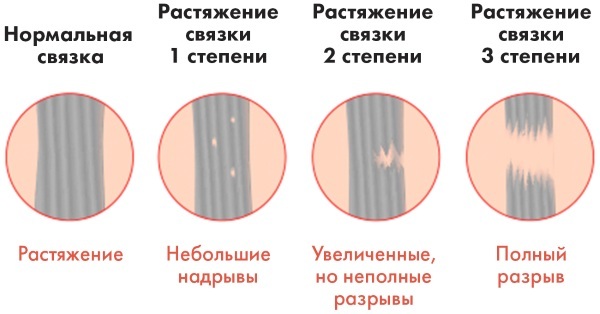
Most chest deformities are grade 2.
Symptoms and Signs
Patients with chest tightness experience sudden pain, a tearing sensation in the chest or front of the shoulder during vigorous work or sports activities. With minor tears, pain, stiffness occurs after rest or later (especially at night or the next morning). In severe cases, pain is incapacitating, preventing the patient from taking further action.
- The pain is localized to the chest and front of the shoulder or armpit.
- Sometimes it radiates to the shoulder or neck, increases with physical activity.
- Patients usually experience pain when the affected pectoralis muscle is strongly touched and often when trying to stretch the muscles or perform actions that require a strong contraction.
- Hematomas and swelling of the chest. Muscle injury causes bleeding from the torn muscle mass, blood collecting in the subcutaneous tissue, leading to a hematoma. A tendon rupture causes less bleeding than a muscle rupture. A bruise is observed over the torn muscles.
- Weakness of muscles. Loss of connection between the torn muscle segment or ligament causes a decrease in muscle strength.
Patients with a minor rupture may have little or no symptoms. In severe cases, a hole (or deformation) in the muscle is found at the rupture site by palpation.
Causes
Stretching of the chest often occurs suddenly due to the fact that a lot of force is passing through the muscle and tendon, which cannot withstand the load. This usually happens during strength training, especially when doing bench press, from the chest.
In other cases, chest tension develops from repetitive or prolonged actions that strain the muscles. This causes their gradual degeneration, weakening, which predisposes to further injuries.
Spontaneous rupture is rare but occurs in older patients. Injury occurs as a result of abrupt activation of the muscle after prolonged inactivity, which led to muscle atrophy.
Chronic corticosteroid use can rupture the pectoralis major muscle, as drug use tends to weaken tendons and muscles.
Treatment methods
Initial treatment includes ice application and immobilization of the shoulder, arm, and chest. With a complete rupture of the tendon of the pectoral muscle, surgical intervention is possible.
| Treatment methods | The ways |
| Conservative | A cold compress prevents further bleeding by causing blood vessels to shrink, reduce swelling and pain. The compress is left on the injured muscle for 30 minutes. and repeated every 4 hours. |
| Hand movement is limited to bandaging around the chest and limb. | |
NSAIDs are used for severe pain: Diclofenac, Meloxicam IM. Muscle relaxants used to relieve muscle spasm: Baclofen, Skelaxin, Flexeril. | |
| Exercise therapy and physiotherapy | Physical therapy for the first 8 weeks is recommended passive. Starting from 11-12 weeks, you can exercise using a force of 2 to 5 kg. Physiotherapy is helpful in post-operative rehabilitation. Soft tissue massage. |
| Surgical | In cases of complete rupture of the pectoral muscle, surgery may be indicated. |
After conservative treatment, the strength of internal rotation and adduction is not fully restored. It is only possible to achieve the full amplitude of painless movements, which is sufficient to restore full-fledged daily activity. Athletes wishing to return to exercise may require surgery.
Poland syndrome
It is a rare birth defect characterized by the absence or abnormal structure of the pectoral muscle. Often accompanied by other anomalies.
A congenital disease can affect anyone. For some unknown reason, the right side of the body is affected twice as often as the left. Boys are affected 3 times more often than girls.
Classification
It is currently classified as a "nonspecific developmental defect" that occurs around the sixth week of fetal development.
There are 4 types of chest formation in this pathology:
- there are no violations of the cartilaginous and bone part of the ribs, the anomaly manifests itself at the level of soft tissues;
- the bone and cartilaginous parts of the ribs are preserved, but the chest is not developed correctly;
- there is hypoplasia of the costal cartilage, but the bony parts of the ribs are preserved, while there is a slight deformation of the sternum;
- lack of cartilage and bone tissue at 2-4 ribs.
Symptoms and Signs
Abnormal signs are visible even to non-specialists and are detected by parents in the first days after the birth of the child.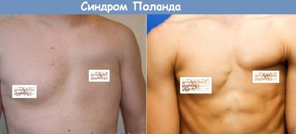
Frequent signs:
- Lack of pectoral muscles.
- Gastrointestinal disorders. liver and biliary tract.
- Monkey crease on the affected side: the only line through the palm. There are usually three folds on the palms.
- Brachydactyly or short fingers.
- Abnormal ribs.
- Asymmetrical upper limbs.
- Diaphragm defects.
- Absence or abnormality of the ulna and radius.
- Syndactyly or webbing, fusion or joining of the fingers together.
- Oligodactyly or missing fingers.
- Missing or underdeveloped nipples.
- Pathology of the scapula or humerus.
Causes
The exact cause of the syndrome is unknown, but specialists in thoracic surgery suggest that the cause may be a violation of the migration of embryonic tissues necessary for the formation of the pectoral muscles.
And also many believe that:
- the syndrome is sporadic (not inherited) because it occurs in people who do not have a family history of the syndrome;
- in some cases, this occurs in an autosomal dominant manner. Children who do not inherit the abnormal gene will not develop the disease and pass it on to their offspring.
Treatment methods
The most acceptable treatment for the syndrome is reconstructive surgery, which includes:
- rebuilding the chest using existing chest muscles taken from other parts of the body;
- patients with slight chest asymmetry are treated with a permanent implant expander;
- deformities of the fingers can be corrected by surgical reconstruction and detachment of the fused or webbed fingers:
- surgical treatment of diaphragmatic hernia.
For boys, reconstructive surgery can be done from the age of 12, and for girls, it is postponed until the breasts are fully developed. After surgery, physiotherapy is used to improve the child's condition and treat muscle weakness
The pectoralis major muscle is a dynamic and multifunctional muscle. Knowledge about the structure of the organ, the places of its attachment to parts of the skeleton, helps to increase awareness of the body, the permissible range of physical exercises.
Video about the pectoralis major muscle
Pectoralis major anatomy: