Content
- What is trisomy
- The rate of indicator in pregnant women in the first trimester
- Indicators of deviations
- The main causes of chromosomal mutation
- What varieties are there?
- Trisomy 13, Patau syndrome
- Causes
- Signs
- What do newborns look like?
- Forecast for children
- Trisomy 18, Edwards syndrome
- Causes
- Signs
- What do newborns look like?
- Forecast for children
- Trisomy 21, Down syndrome
- Causes
- Signs
- What do newborns look like?
- Forecast for children
- Can a chromosomal mutation be cured?
- Trisomy 21 video
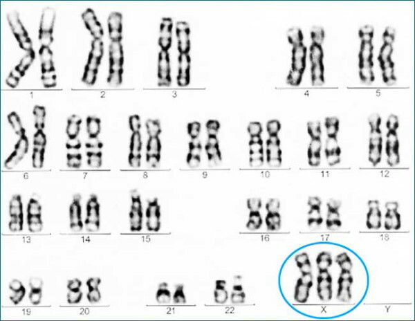
Trisomy is a genetic mutation, the development of which is due to abnormalities in chromosome pairs 21, 18 or 13. The pathology is congenital and is detected during a routine ultrasound scan during pregnancy.
What is trisomy
Pathology is a malfunction in the formation of chromosomal pairs. In this case, both an extra pair of chromosomes and their deficiency can be observed. As a result of such violations, genetic abnormalities develop, which have various manifestations and causes of occurrence.
The rate of indicator in pregnant women in the first trimester
To identify genetic disorders, pregnant women need to systematically undergo ultrasound diagnostics. The study allows you to determine the presence of anomalies at an early stage.
During the examination, phonometric indicators are assessed. In this case, the main criterion is to determine the thickness of the collar space (TVP). Its norms are 0.7-0.8 mm.
The structure of the facial structures, the skull is also studied, the parameters and the presence of the nasal bone are determined (it can be detected already by the 10th week of pregnancy). Closer to the 12th week, its dimensions are from 2 to 3 mm.
Additionally, fetal heart rate measurements are taken, blood flow in the venous duct is assessed, since violations in it may indicate the development of a pathology such as Down's syndrome.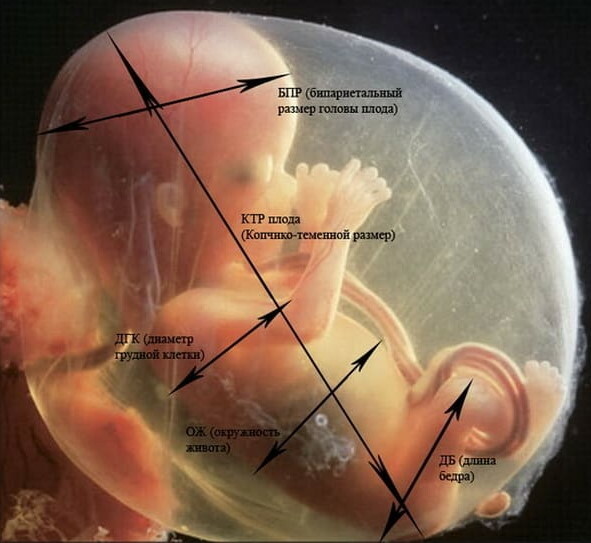
The defining values are the parameters of the coccygeal-parietal and biparietal regions:
| Index | Gestational age | Norm |
| Coccyx-parietal size | 11 week | 34-42 mm |
| 12 week | 42-51 mm | |
| 13 week | 51-63 mm | |
| Nasal bone | 11 week | Considered but not measured |
| 12 week | 2-3.1 mm | |
| 13 week | 2-3.1 mm | |
| Heart rate (HR) / min. | 11 week | 153-165 |
| 12 week | 150-162 | |
| 13 week | 147-159 | |
| BPR (biparietal size) | 11 week | 13-17 mm |
| 12 week | 18-21 mm | |
| 13 week | 20-24 mm | |
| LZR (frontal-occipital size) | 11 week | 19-21 mm |
| 12 week | 22-24 mm | |
| 13 week | 26-29 mm | |
| HCG (ng / ml) | 11 week | 17,4-130,4 |
| 12 week | 13,4-128,5 | |
| 13 week | 14,2-114,7 | |
| PAPP-A (honey / l) | 11 week | 0,46-3,73 |
| 12 week | 0,79-4,76 | |
| 13 week | 1,03-6,01 |
Indicators of deviations
Trisomy 21, 18, 13 is determined using various diagnostic methods, therefore, their passage during pregnancy is considered mandatory.
You can determine the presence of abnormalities using an ultrasound scan. However, diagnosis in the first weeks of pregnancy is ineffective. In this connection, it is recommended to undergo other research methods.
The most informative and often prescribed tests are blood tests and fetal biopsies.
The blood is drawn from the mother. When evaluating the indicators, the accepted norms are taken into account. The condition can be assessed by indicators such as pregnancy-associated plasma protein A (PAPP-A) and human chorionic gonadotropin (hCG).
Low levels of PAPP-A and hCG clearly indicate one of the chromosomal abnormalities. An increased level of indicators indicates problems with bearing a child.
The main causes of chromosomal mutation
At the moment, the exact reasons for the development of violations are being studied. However, experts identify several predisposing factors that increase the chances of having children with chromosomal abnormalities.
Possible reasons:
- age over 30 (for women);
- age over 40 (for men);
- hereditary factor (in the genus there are relatives with genetic disorders in the chromosomal series);
- problems during a previous pregnancy - missed pregnancies, miscarriages, abortions;
- infectious diseases that have arisen during the period of conception of a child or directly in the first stages of pregnancy.
An altered number of chromosomes (aneuplodia) is observed in children born to women over 35 years old. At the same time, the risks of developing such violations are 1%.
What varieties are there?
Pathology is classified according to the affected pair of chromosomes. Each type of violation has its own name, manifestations, characteristics and other distinctive features.
Trisomy 13, Patau syndrome
This disorder is rare and occurs with the same frequency in both sexes. At the same time, the risks of developing the syndrome increase with the age of the parents. It is possible to determine the presence of the disease from the 9th week of pregnancy using ultrasound diagnostics or a blood test.
Causes
Trisomy 21, 18, 13 can occur as a result of various provoking factors, which is almost impossible to establish exactly.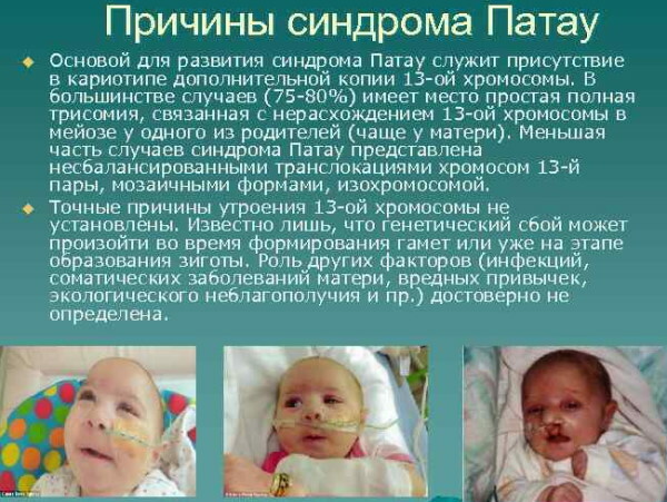
At the moment, the exact origin of the anomaly is unknown. Most experts are inclined to the version of an accidental genetic disorder in chromosomal pairs. Pathology develops as a result of the formation of an additional chromosome in the 13th pair. The influence of somatic disorders, infectious diseases, bad habits and other factors has not been established.
Signs
The symptoms of pathology are diverse and affect many different systems and internal organs.
Signs:
- small body weight - up to 2.5 kg;
- asphyxia (suffocation);
- abnormal development of the brain;
- defects in the structure of the face and skull;
- central nervous system disorders - hydrocephalus, cerebellar underdevelopment, cataracts, deafness, optic nerve damage;
- congenital heart defects;
- focal absence of hair or skin;
- altered foot structure - absence or presence of toes;
- abnormalities in the urinary system;
- malformations of the digestive tract;
- underdevelopment of the musculoskeletal system;
- abnormal structure of the reproductive and reproductive system.
Children with Patou syndrome are developmentally delayed (mental and physical retardation).
What do newborns look like?
You can determine the pathology by the appearance of the baby. Children with the disease are characterized by a small head circumference, a low and sloping forehead, and narrow eye holes. The bridge of the nose has a flat shape and sinks slightly into the interior of the skull.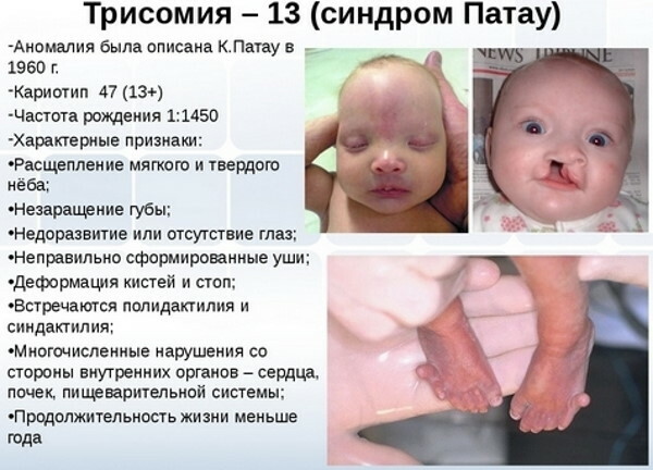
At the same time, the development of other facial defects (cleft palate, cleft lip) is also noted. The auricles are also prone to deformation and are located lower than in healthy children.
Forecast for children
The prognosis for the disease is unfavorable. More than 95% of all newborns die in the first months after birth. The remaining percentage of children have severe psychomotor developmental disorders, including complex forms of idiocy.
Trisomy 18, Edwards syndrome
Trisomy 18 differs from abnormalities in chromosome pairs 21 and 13. Pathology ranks second among chromosomal abnormalities and is characterized by abnormalities in the structure of various internal organs and systems.
According to statistics, most girls are susceptible to the disease.
Pathology is divided into 3 main forms - complete, partial and mosaic.
Causes
Pathology occurs in the case of the formation of an additional pair of chromosomes. This factor can be influenced by various reasons. At the same time, a relationship was established between the frequency of development of the disorder and the age of the mother - the older the woman, the greater the risks.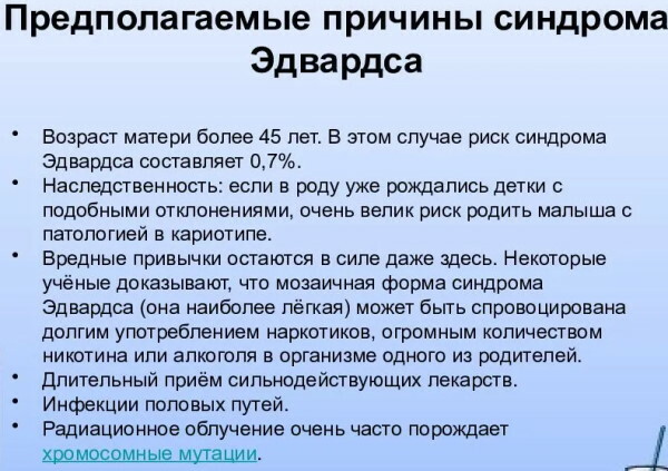
Possible factors:
- the mother's age is over 40;
- bad habits (smoking and alcohol);
- taking medications (especially in the 1st trimester of pregnancy);
- infectious diseases of the reproductive system (research has not been conducted);
- radiation radiation.
In some cases, the disease develops as a result of a genetic predisposition.
These factors are only an assumption, since at the moment the exact causes of the pathology are unknown. It is believed that when a child is born with Edwards syndrome, the risk of having another child with similar abnormalities is 2-3%.
Signs
With this pathology, pregnancy is impaired. During the gestation of a child, there is low water, low fetal activity, small size of the placenta.
After birth, the child himself has various anomalies in the structure of systems and internal organs.
Signs:
- low body weight (up to 2170 g);
- asphyxia;
- congenital heart defects;
- pathology of the gastrointestinal tract (hernia, fistula, atresia);
- abnormalities in the genitourinary system;
- malformations of the central nervous system - hydrocephalus, hypoplasia, cysts;
- problems with swallowing, sucking, breathing;
- mental retardation, imbecility, idiocy.
In most cases, additional disorders develop due to complications of the pathology.
What do newborns look like?
External signs of the disease are characterized by abnormalities in the structure of the head and limbs. Skin signs also predominate.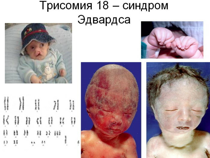
External signs:
- dolichocephaly - long and narrow skull;
- microcephaly - a discrepancy between the size of the head and the body (the size of the skull is too small);
- low location of the ears - cartilage reliefs are little or completely absent, narrowing of the ear canal, absence of a lobe and nodule;
- non-closure of the soft or hard palate, lips;
- changes in the foot and toes - abnormal length of the toes, clubfoot, fusion of fingers;
- underdevelopment of the penis and hypertrophic changes in the clitoris;
- flexor position of the hands.
Also, a child with this syndrome has inadequate emotional reactions, sometimes up to their complete absence.
Forecast for children
The prognosis for pathology is almost always unfavorable. About 55% of children with abnormalities do not live to be 3 months old. About 10% of newborns survive to 1 year.
Children who live to an older age have health problems and need constant care. To prolong the life of a child, it is necessary to carry out complex operations on the heart, kidneys or other organs. The chances of a full and long life in children with Edwards syndrome are virtually nonexistent.
Trisomy 21, Down syndrome
Trisomy 21, 18 and 13 are diagnosed in most cases in children born to parents over 40 years of age. Among the most common genetic disorders, the first place is taken by the pathology of the 21st chromosome series.
Down syndrome is a genetic disorder that occurs in 1 child out of 700 infants.
Pathology occurs as a result of nondisjunction of chromosomes in the male reproductive cell. However, according to statistics, an additional chromosome is transmitted from a woman in 88% of cases, from a father in only 8%. In other cases, the anomaly occurs as a result of improper cell division after fertilization.
Causes
At the moment, there are only 2 main reasons for the development of the disease. The first of these is the age of the parents. The larger it is, the higher the risk of the syndrome.
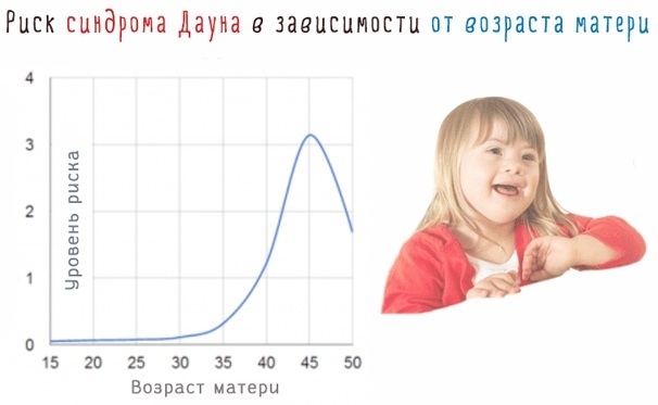
For parents aged 35-40 years, the risk of having a child with a genetic abnormality is 1 in 1000. After 40 years, these risks increase significantly and amount to 1 in 60.
The second reason for the development of the disorder is heredity. If the family has or had relatives with similar pathologies, the risks of developing this disease increase significantly. Also, the disease can occur in children born from marriage with blood relatives.
It is believed that factors such as poor environmental conditions, bad habits, radiation do not affect the birth of children with Down syndrome. The exact mechanisms and reasons are currently being established.
Signs
Children with a genetic disorder have characteristic external signs. It is possible to determine the presence of pathology even at the stages of bearing a child during an ultrasound scan.
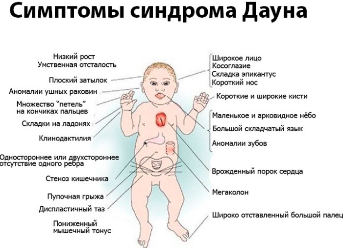
Symptoms:
- disproportionate size of the head in comparison with the rest of the body (the size of the skull is too small);
- low body weight at full gestation;
- delay in physical, mental and mental development;
- cardiac disorders - heart defects (heart failure), shortness of breath, hyperhidrosis, the presence of noise;
- disorders of the digestive tract - intestinal obstruction, dyspeptic disorders, obstructive disorders in the duodenum;
- concomitant diseases - eye diseases, hearing problems, skin manifestations;
- tendency to obesity, the occurrence of Alzheimer's disease, the development of infertility.
 In the first days and years of life in children with pathology, various respiratory and inflammatory diseases are found, which is why, in the absence of timely assistance, they die.
What do newborns look like?
Trisomy 21, 18, 13 has similar external and internal signs, therefore, additional studies are required to establish an accurate diagnosis. Outwardly, children with Down syndrome have significant differences from healthy children. Thus, they have various defects in the structure of the body, limbs, and head.
External signs:
- in profile, the child's face is flat;
- microcephaly (not observed in all cases);
- flat nose;
- the formation of skin stores around the back of the neck;
- narrow incision of the palpebral fissures (Mongoloid type);
- Brushfield spots (white blotches on the iris of the eyes);
- open mouth;
- protruding tongue;
- monkey fold on the palms;
- shortened fingers;
- the presence of 2 phalanges on the fifth toe.
A shortened neck, small ears, and wide hands are also noted.
Children have hypermobility (joint mobility). This is due to the underdevelopment of the connective tissue.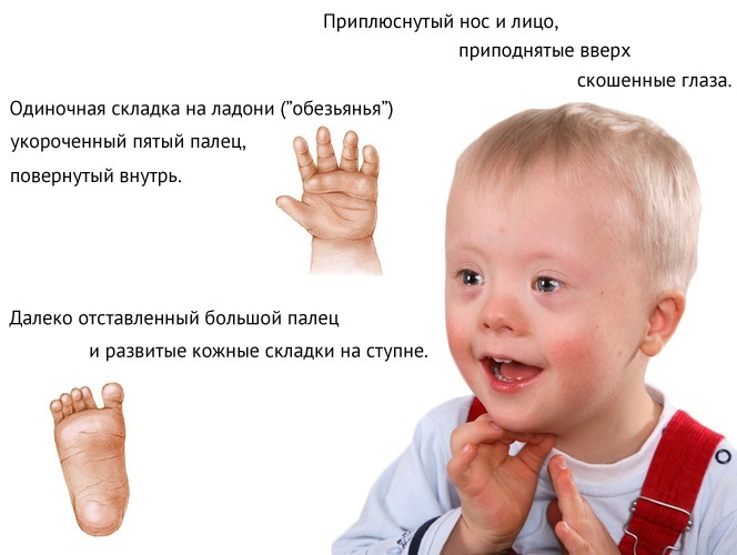
In rare cases, some newborns show signs of chest and spinal deformities.
Forecast for children
With a disease, the prognosis is the most favorable among all chromosomal abnormalities. This is due to the fact that the violation has not so critical defects and anomalies in the structure of internal organs and systems.
Qualified medical care and care in the first years of a child's life can reduce the risk of mortality, since in this for a period of time, the newborn is susceptible to the development of respiratory diseases, to which a child with Down syndrome is predisposed.
With properly organized child care, life expectancy is 40-45 years.
Can a chromosomal mutation be cured?
Chromosomal abnormalities are not clinically curable. The vast majority of children die in the first months of life. With proper care and medical attention, the chances of a child's life extension are increased. However, constant monitoring, complex operations and care of such patients are required.
Down syndrome is the most dangerous for a child's life. With this disease, the chances of living longer than 30-40 years are several times higher than with other chromosomal abnormalities.
At the moment, the issue of defects in the chromosome series is being studied by specialists, but effective methods of treating pathologies have not yet been developed.
Trisomy in the 18th, 21st or 13th chromosome row is a life-threatening disorder for a child, therefore, before conception and during pregnancy, the future the mother should take into account all the risks of the development of pathology and undergo systematic studies to identify signs of abnormalities in the early stages.
Trisomy 21 video
Down Syndrome. Trisomy 21. Ultrasound: