 Very often, especially in childhood, you have to hear a terrible diagnosis - exostosis. What is this disease, and is it so dangerous?
This is an bone-cartilaginous or bony proliferation of a non-tumor nature on the surface of the bone .First the neoplasm consists only of cartilaginous tissue, but eventually it hardens and is transformed into a spongy bone.
Above is a cartilage coating a few millimeters thick. It then serves as the basis for the further growth of the tumor.
The main danger of the disease is that it develops very slowly and is asymptomatic. The size of the growths can range from a few millimeters to ten or more centimeters.
Another feature of exostosis is that it is usually diagnosed in adolescence, when there is an intensive growth of the skeleton. There is also a theory about a hereditary predisposition to the disease, but it is not confirmed.
Contents of the article
- Causes and Risk Factors
- Features of the cartilage growth
- Classification and localization
- Symptoms and Diagnosis
- Removal of accretions
- Recovery after surgery
- Complications of the disease
- Preventative measures
- Instead of withdrawal
Causes and risk factors
Growth formation occurs for various reasonsand depends on many factors.
It can be:
- injury or infringement;
- dysfunction of the endocrine system;
- abnormalities in the development of cartilage and periosteum;
- is an inflammatory process;
- some infectious diseases( for example, syphilis).
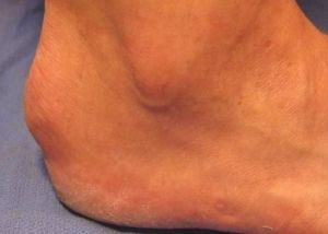
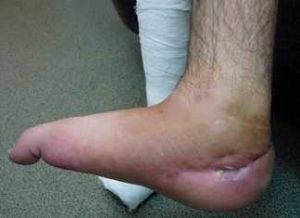
On the photo exostosis calcaneus
Today, a large number of studies are aimed at studying the heredity of this disease.
However, despite the fact that many cases of family exostoses are known, most scientists are skeptical about this theory. After all, it does not explain single cases of the disease, and therefore can not be the only true one.
Settling on the bones, this element eventually leads to the formation of outgrowths. Hypercalcemia can occur due to excessive consumption of eggs, dairy products, cabbage, parsley, or because of hard water.
Features of the cartilaginous outgrowth
Bone-cartilaginous exostosis, or osteochondrosis is a benign bone tumor, formed from cartilaginous tissue.
The disease, as a rule, does not appear until 8 years, but during the period of active growth of the skeleton - from 8 to 17 years - the probability of its development increases several times. Most often it is diagnosed in adolescents during puberty.
With osteochondrosis, the number of growths can vary from a few to dozens.
On this basis, the disease is divided into two types:
- The solitary bone-cartilaginous exostosis. Always represented by one tumor. It can be of different sizes and is fixed. With a significant increase in the tumor can put pressure on the vessels and nerve trunks;
- Multiple exostosis chondrodysplasia .This type of disease is characterized by the appearance of several neoplasms. Chondrodysplasia is most often inherited.
Classification and localization of
In most cases, exostosis is diagnosed on the shoulder joint, hip, collarbone, scapula, tibia.
According to statistical data, 50% of all exostoses are attached to the tibia and femur. Significantly, the disease affects the hands and feet. Also, there are no known cases of formation of growths on the skull.
If the disease affects the spine, then with its further development, spinal cord compression may occur.
Symptoms and Diagnosis
 The disease develops very slowly and, as a rule, is asymptomatic. It may take years before the disease is detected. The only exception is when the growths press on vessels or nerve endings.
Then there may be pain in the area of compression, a feeling of numbness or goosebumps, headaches, dizziness.
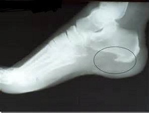
Most often, the disease is detected by chance during the X-ray examination. Without x-ray diagnostics is practically impossible.
Carrying out this type of research allows you to tell about the number and shape of neoplasms, their size and development. At the same time, it must be taken into account that the cartilaginous coating that covers the growth is not visible in the picture.
Therefore, the actual size of the tumor is always greater than it seems.
Removal of
build-up. There is no method of conservative treatment of the disease. If necessary, the enlarged areas of bone tissue are removed during the surgical procedure.
Children under 18 years of age try not to carry out operations in view of the fact that independent resolution of exostoses is possible.
The operation is carried out:
- if on face rapid proliferation of tissues;
- if the tumor is so large that it is released on the surface;
- if the growths squeeze the vessels or nerves.
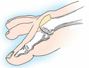
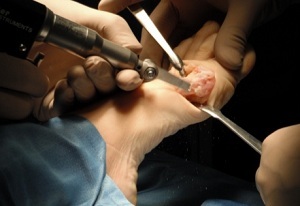
Operative treatment is performed under local or general anesthesia, depending on the location and size of the lesion. First, using a chisel, the bone outgrowth is removed, and then bone is smoothed out with special tools.
On video removal of exostosis of the auditory meatus:
Recovery after operation
Rehabilitation takes no more than two weeks. If only one tumor was removed, the patient could get out of bed the next day.
Recovery after the operation is divided into two stages. On the first, a gentle motor regime is established. Then, when the edema decreases, a restorative regimen is prescribed. In the postoperative period, it is very important to return the muscles to their strength.
It is necessary to achieve the condition that the training exercises do not cause pain. Only then the restoration is considered successful.
Complications of the disease
In most cases, exostosis does not carry a great danger, but sometimes complications of the disease occur. It is worthwhile to worry if the growths are formed in the region of the spine.
Then, with intensive growth, they can squeeze the spinal cord, which leads to serious consequences.
In children and adolescents with the development of multiple chondrodysplasia, deformations of the skeleton are likely. Sometimes, though rarely enough, such a pathology is diagnosed as a fracture of the exostosis leg.
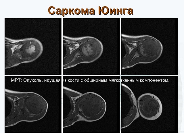
If neoplasms start to grow quickly, there is a possibility of their malignant degeneration.
As a rule, cancerous tumors are formed on the thigh, vertebrae, scapula, pelvis. They can have a morphological structure of spindle cell sarcoma, chondrosarcoma and other species.
Preventive measures
 To date, there is no specific system of preventive measures for this disease.
The only way to prevent accretions is regular examination and examination. Especially important is this prophylaxis for children, since they have bone growths that can cause deformation of the skeleton.
In addition, it is always necessary to have a prophylactic examination after an injury. Any bruise, damage to the nails or fracture of the bone can lead to the development of the disease.
Instead of output
Whatever the reason for the development of exostosis, it is not worth to be afraid of. In fact, the disease is not as terrible as it might seem at first.
Yes, in some cases, with intensive growth of a tumor, it can indeed degenerate into a malignant tumor. However, this happens rarely enough.
In most cases, the prognosis for life in this disease is favorable. Bony growths are successfully removed in any clinic without any consequences. And sometimes even an independent resolution of the disease is observed.
This happens in children, when the disease passes spontaneously. So do not panic. Believe in the best - and the disease will necessarily recede.