Indications for ECHO of the heart there are good reasons. The procedure may be prescribed if the patient complains of pain or murmurs are diagnosed on auscultation. Another common cause is high blood pressure.
Record content:
- 1 Method description
-
2 Classification
- 2.1 Doppler
- 2.2 Transesophageal
- 2.3 Stress
- 3 Indications
- 4 Possible contraindications and limitations
- 5 What does it show?
- 6 Preparation rules
- 7 Stages
- 8 Decoding the results
- 9 Possible complications
- 10 Conducting frequency
- 11 Video about ultrasound of the heart
Method description
Echocardiography is a simple and painless medical procedure that shows possible problems in the functioning of the heart. The essence of echocardiography is the use of ultrasound - high-frequency waves that are not captured by our hearing organs.
By means of a special sensor attached to the patient's body, the waves penetrate into the tissues, as a result of which their main characteristics - frequency and amplitude - change. These changes depend on the state of the organs. Waves with altered characteristics return to the sensor, where they are converted into an electrical signal.
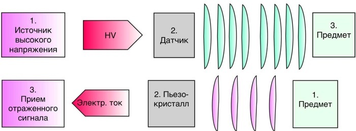
The next step is echocardiographic processing. As a result, a complete picture of the study of the organ from all sides is formed. On the monitor, the doctor sees a flat or three-dimensional image, which, if necessary, can be transferred to a sheet of paper.
For the first time, ultrasound waves to determine the state of the heart were used by Swedish scientists in the 50s. last century. They created a special device with which it was possible to receive signals from the heart ventricle and one of the valves.
More than half a century has passed since the creation of the device. Since then, it has been modernized, and ultrasound diagnostics of the heart has improved with it.
Classification
The procedure can be carried out by one of several methods. The main difference between the two is the way the signal is played. This can be 1 of 3 types of modes.
Each mode has a distinctive letter in its name:
| Mode | Description |
| BUT | The letter "A" indicates a relationship with amplitude. A-mode is the registration of signals in the form of peaks. The amplitude is related to the intensity of the signal. |
| M | Moving structures can be seen in M-mode. This regime is based on movement. The letter M is English and stands for the word "motion", which translates as "movement". M-mode is used most often in diagnostics. Together with him, 2-dimensional echocardiography and Doppler echocardiography are performed. When a survey is carried out in M-mode, signaling occurs along a single selected axis. As a result, an image of the heart is formed in the 1st plane. This mode can even be applied to newly born babies. When performing 2-dimensional echocardiography, an image is obtained in 2 planes. Therefore, the specialist conducting this examination has the opportunity to study the movement of the structures of the heart and analyze it. |
| IN | In this mode, the intensity of the signals is demonstrated by the brightness of the luminescence. The mode got its name due to the initial letter of the English word "brightness", which means "brightness" in Russian. |
Doppler
Through a procedure called Doppler echocardiography, blood flow velocity and turbulence can be determined.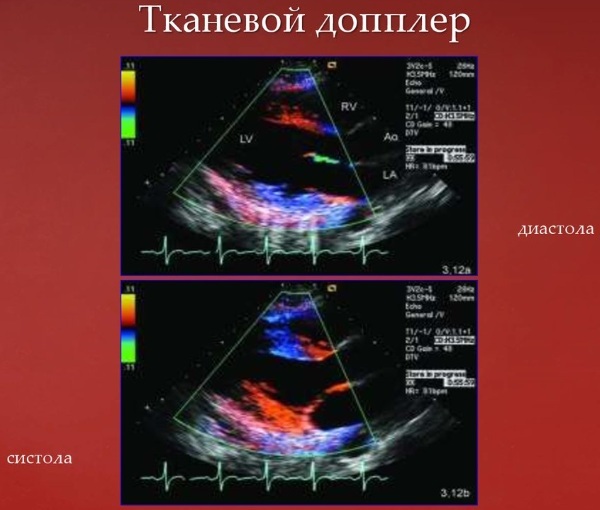 The method is based on the so-called Doppler effect. Its essence is as follows: the speed with which the object moves affects the frequency of the supplied signal.
Transesophageal
The named and described above examination methods are transthoracic echocardiography. This procedure is not always possible. It cannot be performed when there are sound barriers preventing the signal from passing.
This obstacle, for example, can be subcutaneous fat. In this case, another type of echocardiography is used, which is called transesophageal. The essence of this procedure is as follows: first, the patient must swallow the sensor, then the examination is carried out.
This procedure can be additionally performed after conventional echocardiography. In this case, it will allow you to obtain additional data and clarify the diagnosis. This type of examination is of particular relevance when it is required to obtain information about those parts of the heart that are located near the esophagus and at the same time are removed from the surface of the body.
However, it should be borne in mind that this method of echocardiography is contraindicated for those who have problems with the esophagus.
Stress
This echocardiography is used to detect hidden abnormalities that may appear during stress. In addition, this method allows you to identify problems that arise during heavy physical exertion or under the influence of drugs.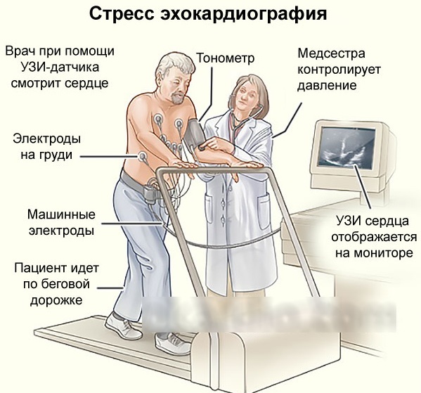
Stress echocardiography requires a minimum of 2-dimensional echocardiography. Also, a prerequisite for such a survey is the presence of several highly qualified specialists - 2 doctors and 1 nurse.
During the survey, various kinds of stress agents are used. These can be special pharmacological agents. Also, the patient may be asked to perform certain movements in order to increase physical activity. For example, you will need to pedal or run. When performing physical exercises, the load will increase and then the specialist will be able to monitor the change in pressure.
First, an analysis of the data of the heart in a normal state is carried out, and then the work of the organ is studied at its maximum load.
Indications
An echo of the heart shows possible problems and is prescribed for such signs as:
- feeling tired, lethargic;
- pain that is localized in the heart region;
- cold hands and feet;
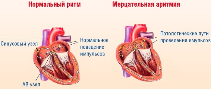
- arrhythmia, including when drinking alcohol;
- frequent blanching of the skin;
- noises that are detected during auscultation;
- heart failure;
- high blood pressure;
- too slow weight gain in children;
- frequent headaches and loss of consciousness;
- high body temperature, accompanied by a rapid heart rate and shortness of breath. It is not associated with other diseases;
- cough.
Also, this procedure may be recommended when there is a suspicion of the presence of fluid in the pericardial region. The most common reason for the appointment of echocardiography is the need to identify a heart defect.
An ultrasound of the heart can be prescribed for:
- ischemic heart diseases;
- problems associated with the heart membranes;
- myocardial infarction. In this case, echocardiography is usually done for prophylaxis;
- changes in the size of the heart or blood vessels, as well as their position and shape, which was revealed as a result of an X-ray examination.
When performing echocardiography using modern devices, many indicators related to heart contractions can be distinguished. The specialist will be able to identify the pathology and prescribe an appropriate therapeutic course, including for the initial stage of the contractile function.
Regular ultrasound examination of the heart is necessary for everyone who is professionally involved in sports.
As for children: cardiography is prescribed for them for the same reasons as for adults. In addition, every adolescent who is 14 years of age must be tested because this age, the body is growing rapidly and there is a high probability of the appearance of pathological changes in a heart.
According to the norms accepted in our country, all children whose birth has passed 1 year should consult a cardiologist, having previously done echocardiography.
ECHO of the heart shows problems in the early stages, therefore, the procedure is recommended for all expectant mothers, because during pregnancy the load on the heart and vascular system increases.
Possible contraindications and limitations
Given that this procedure is safe, it can be performed on a person at any age. Thanks to this method, you can find out about the state of the heart of an unborn child.
The procedure can be repeated many times, since it has no negative consequences. Contraindications also apply to transesophageal examination. This procedure should not be performed for those who have problems with the esophagus.
Also, the following factors can interfere with echocardiography:
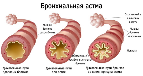
- bronchial asthma;
- long-term smoking;
- severe deformation of the chest;
- large breast size.
What does it show?
With echocardiography, the doctor receives the necessary information about the patient's heart.
The procedure allows:
- to assess the functioning of the heart;
- see its size and their possible deviations from the norm;
- determine how thick the heart walls are;
- measure the pressure not in the vessels, but directly in the heart;
- evaluate blood flow.
It must be remembered: timely echocardiography is an effective preventive measure against many diseases associated with blood vessels and heart. ECHO of the heart will allow you to accurately determine what treatment should be.
Preparation rules
Echocardiography does not require special training.
However, there are some conditions for conducting that must be observed:
- The procedure can only be carried out when the patient is absolutely calm. Otherwise, he will have rapid breathing and an increased heart rate.
- It is important that the rate of respiration and heart rate is not higher than the physiological norm. This means that you should refrain from physical activity before performing the procedure.
- The procedure cannot be carried out in stressful conditions.
- Echocardiography preparation also applies to nutrition. It is better to refuse foods that can affect the work of the heart. First of all, you need to pay attention not to food, but to drinks. It is advisable to refrain from coffee, tea and other invigorating drinks, which contain a significant proportion of caffeine.
If these conditions are met, then thanks to the performed echocardiography, the doctor will be able to obtain the maximum necessary information about the organ under study. If this is not possible, the patient must be sure to inform about what medications he is taking.
Transesophageal echocardiography requires separate preparation. The patient before such a procedure should completely refuse to eat. Abstinence from food should last about 5 hours.
Stress echocardiography also has its own preparation characteristics:
- 3 hours before the start of the examination, you must not give the body a load.
- You should not eat a lot of food before the procedure. Just a couple of hours before the start of the exercise, you can have a light snack and drink a small amount of water.
- The patient should bring light clothing that will not hinder his movement.
Stages
ECHO of the heart shows accurate results, thanks to the use of the latest and most powerful equipment in modern clinics. This greatly simplifies the formulation of the correct diagnosis and the determination of the appropriate method of therapy.
The procedure is performed on an outpatient basis. According to the results of recent studies by specialists, a correctly performed echocardiography procedure does not pose a risk to the patient's health. During the study, the patient does not experience any discomfort. For this reason, it can be carried out not only for adults, but also for children.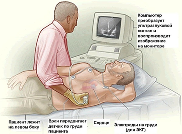
The stages of its implementation are as follows:
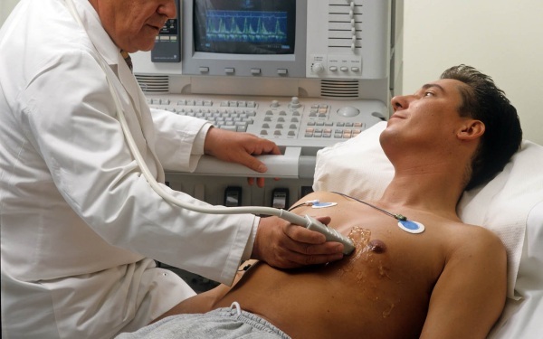
- The patient undresses to the waist. It is important to take care that there are no jewelry and accessories on the body, especially metal ones.
- The patient, stripped to the waist, lies down and completely relaxes.
- A special composition in the form of a gel is applied to the chest.
- A specialist attaches sensors to certain points that must be connected to the equipment.
- On a special screen, you can see the heart and observe its functioning in real time. Thanks to this, possible violations in the work of the organ can be detected.
The average duration of the procedure is 40 minutes. Sometimes it lasts about 20 minutes, and sometimes it can take up to 1 hour.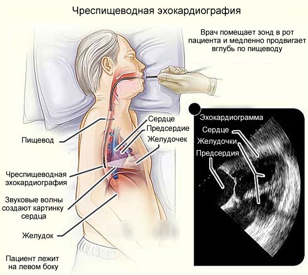
All information that will be obtained during echocardiography is stored in electronic form. The next stage is the interpretation of the data obtained. This is what the doctor does. Subsequently, he must form a conclusion.
Decoding the results
After the examination has been completed, you can see its results. Here, many patients mistakenly believe that the analysis and decoding that were received from the doctor is the finished result. Patients may think that, along with the finished result, they are also given a diagnosis and an established problem associated with the work of the heart. In reality, this is not the case.
The results of the procedure are what was revealed during the study. It is indicated whether there are pathologies and whether the functions of the circulatory system are not impaired. In the event that everything is in order with the heart and blood vessels and no problems have been identified, nothing else will be required. If there are even minor changes, you should definitely contact a cardiologist and consult with him.
The doctor will look at the echocardiography results, talk with the patient, assess the antecedents, diseases and possible causes. Then the correct diagnosis will be made. After that, the doctor is obliged to prescribe the correct therapy that will help improve the condition by eliminating the problem.
After the examination, the analysis of the obtained data begins, the main of which are the parameters of the ventricles of the heart and the partitions separating them.
ECHO of the heart shows:
| Indicators of the right ventricle | Left ventricular indicators |
|
|
The correct analysis of the information received about the heart can only be done by a professional - a cardiologist. If the patient wants to decipher the data about his heart on his own, errors may occur that will entail serious consequences. After all, the data can be influenced by age, as well as physical condition.
Possible complications
Side effects of echocardiography are rare, and severe complications are even rarer. The greatest danger can be carried by transesophageal echocardiography. The study showed that in 7 cases out of 10,000 the heart rhythm was disturbed, in 8 cases hypoxia was observed, in 2 cases bleeding and in 1 case - angina pectoris.
Conducting frequency
The frequency of the examination depends on the results of the 1st procedure. If all indicators were normal, then the next examination can be carried out in 12 months. If some heart problems were identified, then the next procedure should be carried out after 3 months.
Echocardiography is the most reliable way of examining the heart. Thanks to the indications after it, the specialist will receive all the necessary information about the work of the patient's organ.
Video about ultrasound of the heart
Doctor about echocardiography: