
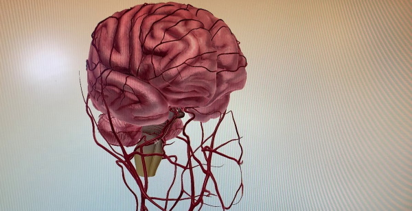
The carotid artery is the main vessel in the human body, supplying blood to the head and neck. Its inner branch supplies oxygen to the brain. Anatomical transformation of a vessel is a reason to undergo a comprehensive diagnosis and urgently begin treatment.
Record content:
-
1 Anatomy, location and structure of the internal carotid artery
- 1.1 Departments
- 1.2 Segments
- 1.3 Branches
-
2 Topography
- 2.1 C1: Cervical segment
- 2.2 C2: Rocky segment
- 2.3 C3: Torn hole segment
- 2.4 C4: Cavernous segment
- 2.5 C5: Wedge-shaped segment
- 2.6 C6: Ophthalmic segment
- 2.7 C7: Communication segment
- 3 What diseases is the internal carotid artery affected by?
- 4 How pathologies are diagnosed
- 5 Methods for the treatment and restoration of the state of the internal carotid artery
- 6 Possible consequences of diseases of the internal carotid artery
- 7 Video about the internal carotid artery
Anatomy, location and structure of the internal carotid artery
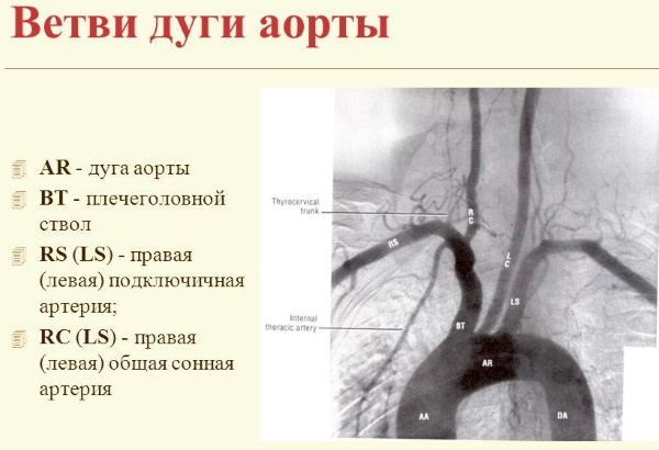
A branched large artery running on each side of the neck is the common carotid artery. It is the main source of oxygenated blood for the head and neck.
This is a paired organ, the left side of which emerges from the aortic arch. The right one departs from the brachiocephalic trunk of the aortic arch. The left vessel is 3 cm longer than the right one, and the length of the right one is from 6 to 12 cm.
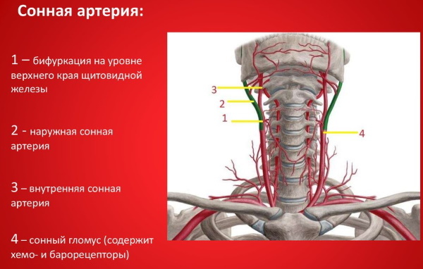
Moving from the sternoclavicular joint, both sides of the common artery go up an oblique path to the upper border of the thyroid cartilage in the cervical region.
At the level of the upper edge, they are divided into 2 arteries:
- internal;
- outdoor.
The internal carotid artery (the anatomy of its parts is visible in the figures later in the article) is larger, designed to supply blood to the anterior structures of the brain, including the hypothalamus and hemispheres.
The artery passes to the side of the pharynx, goes to the base of the skull, enters it through the canal of the temporal bone, perforating the meninges. Then the orbital artery is separated from it, and at the level of the optic nerves, the division into the anterior and middle arteries of the brain occurs. These are the final branches.
The outer one moves up and passes through the parotid salivary gland. In its course, large branches branch off, which supply blood to the structures of the face and neck, including the teeth, gums, and the thyroid gland.
Departments
The common carotid artery ends at the level of the upper border of the thyroid cartilage plate (opposite the disc between the 3rd and 4th cervical vertebrae). At this point, the division into inner and outer parts takes place.
Since the left common vessel departs directly from the aortic arch, it has segments:
- chest;
- cervical.
The right-sided vessel has only the cervical region.
Segments
The internal carotid artery is classified into segments in 2 ways. The newest, proposed in 1998, proposes the division of the artery into 4 parts, the names of which are associated with the regions or anatomical structures through which the interior of the vessel passes.
Namely:
- Stony part (temporal bone).
- Neck part.
- Cavernous part (cavernous sinus).
- Intercranial part.
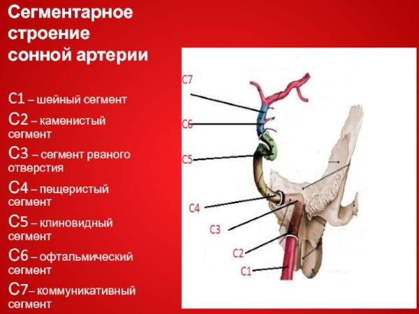
However, in medical practice, many use the older method described in 1996. The names are also associated with the areas through which the artery passes, but they are described in more detail. This classification defines 7 segments.
Branches
The common carotid artery branches into internal and external branches. The inner vessel passes through the cervical, cavernous, stony and cerebral. From the side of the cervical spine, there are no branches.
| Cavernous part | Lower pituitary branches, meningeal branch |
| Stony | Carotidrum branches, arteries leading to the pterygoid canal |
| From the side of the brain | Posterior connective, cerebral anterior, superior pituitary branches, ocular arteries |
The inner part supplies the structures present in the cranial cavity and orbit.
The external carotid artery rises through the upper part of the neck, through the lower jaw, into the parotid gland, where it divides into numerous vessels.
Branches of the external artery:
- upper thyroid, extending to the larynx and thyroid gland;
- lingual to the tongue and sublingual gland;
- facial, passing to the face, palate, tonsils and submandibular gland;
- occipital, going to the sterno-mastoid muscle and the back of the head;
- back, advancing to the auricle, back of the ear and the adjacent part of the head;
- superficial temporal, passing to the head in front of the ear and through the transverse facial ramus to the back of the face;
- maxillary, giving muscle branches to the muscles of chewing;
- meningeal, advancing to the dura mater, teeth, nose, palate and eardrum;
- ascending, which supplies the pharynx, palate, tonsils and dura mater.
The level of changes in the external artery has many variations, which was proved by the Russian surgeon and anatomical scientist N. AND. Pirogov.
Topography
The internal carotid artery (its anatomy is described for informational purposes) runs up the neck and enters the head around the corner of the jawbone, deep to the ear, as an undivided tributary.
This distinguishes it from the external one, which forms several branches on the neck immediately after separation from the common carotid vessel. The topography of the course of the internal carotid artery can be represented using two horizontal S letters. Except for segment C7, odd numbered line segments have no branches.
C1: Cervical segment
This part of the artery runs between the 3rd and 4th vertebrae of the neck (C3 and C4), crosses the transverse processes (bony protrusions) of the upper vertebrae before reaching the carotid canal in the temporal bone at the base skull.
C2: Rocky segment
It is located inside the temporal bone.
Passes from the torn hole, consists of 3 parts:
- ascending;
- bending;
- horizontal part.
It enters the vault of the skull through the lacerated opening of the skull. Arterial branches are usually not visible angiographically, as they can be enlarged when the carotid artery is occluded. The segment supplies blood to the nose and ear.
C3: Torn hole segment
This short segment runs along the cartilage covering the laceration, ending in the petrolingual region. It does not form branches.
C4: Cavernous segment
The cavernous segment C4 forms many small tributaries. These vessels supply structures such as the dura mater and the pituitary gland. They pass through the cavernous sinus with Abducens Nerve.
Branches supply blood:
- ganglia of the trigeminal nerve;
- pituitary;
- meninges.
C5: Wedge-shaped segment
After exiting the cavernous sinus, the internal carotid vessel passes from the proximal to the distal dural ring. There are no designated branches in this segment, although scientists suggest that the ophthalmic arterial branch extends from this segment.
C6: Ophthalmic segment
After passing through the distal dural ring, the artery descends below where it runs parallel to the optic nerve. The branches supply blood to the pituitary gland, eyes, orbit, and nose.
C7: Communication segment
The last segment gives rise to the anterior choroidal and posterior communicating arteries before dividing into the anterior and middle cerebral arteries.
The last 2 arteries, C6 and C7, are terminal branches that enter the brain, which means that the internal carotid artery ends where it branches into the anterior and middle cerebral arteries.
What diseases is the internal carotid artery affected by?
The internal carotid artery (the anatomy of the structure of its walls is associated with the conditions of their functioning), in which a cholesterol plaque has formed, hardens, the lumen narrows in it. It is the main cause of stroke when the flow of oxygen-rich blood to organs and parts of the body is restricted.
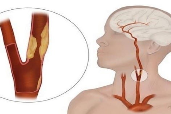
Atherosclerosis can affect any artery in the body. For example, if plaque forms in the coronary (heart) arteries, a heart attack can occur. If in the carotid arteries, a stroke may occur.
A stroke also occurs when blood clots form in the carotid arteries from a ruptured plaque. Fragments of blood cells, called platelets, adhere to the injury, clump together to form clumps that partially or completely block the carotid artery.
A blood clot can also break off the wall of the carotid artery, travel through the bloodstream, and become stuck in one of the smaller arteries in the brain. This blocks the movement of blood, causing a stroke.
The internal carotid artery (the anatomy of the vasculature of the brain is a topical topic for study in medicine) can initially be blocked without noticeable symptoms. For some people, the first sign of illness is a stroke.
The most common diseases of the carotid arteries are:
- Vasculitis - inflammation of the artery due to an autoimmune condition or infection.
- Stroke - A sudden blood clot that interrupts blood flow to the brain causes a stroke. Fragments of cholesterol plaque in the carotid artery can also travel to the brain and cause a stroke.
- Stenosis Narrowing of the carotid artery, usually due to a build-up of cholesterol plaques or atherosclerosis. The disease usually does not cause symptoms until it becomes severe.
- Aneurysm carotid artery. In this case, the weak section of the vessel is inflated like a balloon with each heartbeat. Aneurysms carry the risk of rupture of the vessel, which can lead to stroke or severe bleeding.
- Embolism. In this case, a fragment of a cholesterol plaque or embolus breaks off from the vessel wall, enters the brain, causing a stroke.
- Atherosclerosis carotid artery. Cholesterol plaque, accumulating in the vessel wall, narrows it, causing stenosis, leading to stroke.
- Amaurosis fugax. Temporary blindness in one eye, usually caused by a piece of cholesterol plaque or emboli that has broken off from the vessel wall. The clot can become lodged in an artery that supplies the eye, blocking the movement of blood.
-
Temporal arteritis. An autoimmune disorder in which the branches of the carotid artery become inflamed, known as vasculitis. Symptoms may include fever, severe headache on one side of the head, and jaw pain when chewing.
- Carotid hypersensitivity syndrome. In some people, pressure on the carotid sinus can cause fainting due to a sudden drop in blood pressure. Symptoms can occur while shaving or wearing a tight shirt collar.
Hypoperfusion of any organ leads to a decrease in the supply of oxygen and nutrients to these tissues. Consequently, if tissues remain hypoperfused for an extended period, they die - a process known as a heart attack occurs.
Because the internal carotid artery contains arterial blood, it is under high pressure from the heartbeat.
If an arteriovenous fistula is present, this arterial blood will flow into the low-pressure system of the cavernous sinus and cause blood to flow retrograde into its venous tributaries. These tributaries include ophthalmic veins that drain the orbit and its contents.
If the pressure inside them increases, the patient's eyeball bulges out, and the conjunctiva fills with blood. In addition, the overcrowded cavernous sinus can exert pressure on the structures passing through it, such as cranial nerves III, IV, V1, V2, and VI, eventually causing distinct clinical signs.
How pathologies are diagnosed
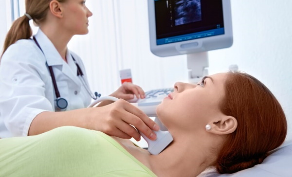
Doctors use one or more visual tests to look inside the carotid arteries and check for narrowing.
Imaging tests include:
- Ultrasound procedure The carotid artery uses sound waves to create images of the inside of a vessel. This is the most common form of imaging for carotid artery disease, and in most cases it detects any narrowing of the arteries.
- Angiography Is an imaging test that uses a special dye. With its help, the vessels are clearly visible in the image.
-
Duplex scanning carotid artery.
- Auscultation (listening to internal body sounds with a stethoscope) carotid arteries.
Methods for the treatment and restoration of the state of the internal carotid artery
Depending on the course of the disease, different methods of treating carotid artery stenosis are selected.
If the carotid artery is narrowed less than 80% and there are no symptoms, then in this case, rapid progression stenosis can be prevented with cholesterol-lowering drugs and aggregation inhibitors platelets.
Doctors recommend the following groups of drugs:
- antihypertensive drugsa, lowering blood pressure (hydrochlorothiazide + Losartan, Losartan, Lorista);
- lipid-lowering drugs, lowering the level of lipids in the blood (Atorvastatin, Rosuvastatin);
- antiplatelet drugs (Aspirin-cardio, Ilomedin, Cardiomagnet, Clopidogrel).
If the carotid artery is narrowed by 80% or more and is not causing symptoms, surgery is advisable.
Possible treatments include:
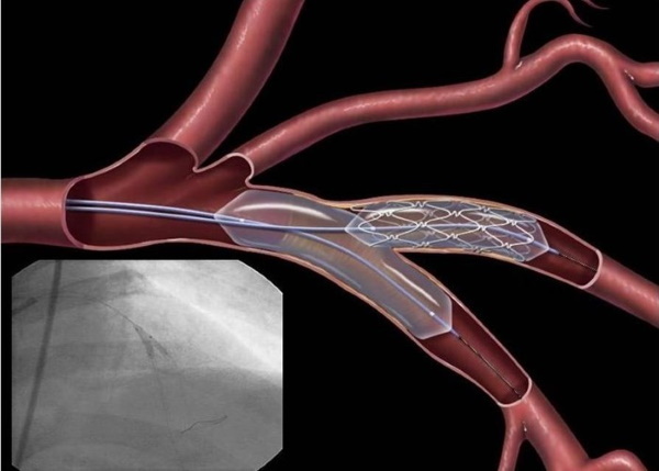
-
Angioplasty and stenting dilate the carotid arteries and increase blood flow to the brain if a person has severe narrowing due to plaque on the vessel walls. During an angioplasty procedure, doctors insert a thin tube with a tiny deflated balloon at the end into a narrow artery. Once the tube is in place, they inflate the balloon, which pushes plaque against the artery wall, helping to restore blood flow through the artery. After the artery has widened, doctors place a stent in this area - a thin mesh tube that will support and strengthen the artery from the inside out and prevent the artery from narrowing again in this area.
- Carotid endarterectomy - The most common form of surgery for severe carotid artery disease. After local or general anesthesia, the doctor makes an incision in the front of the neck, opens the carotid artery, and removes the blockage. This procedure can have a long lasting effect and prevent subsequent strokes.
Possible consequences of diseases of the internal carotid artery
Stroke is a major potential complication of arterial disease. It occurs when blood flow to the brain stops. This can lead to the loss of its functions or even death.
Carotid artery disease causes stroke in several ways:
- Narrowed carotid arteries stop supplying the brain with enough blood.
- A piece of hardened plaque can break off and become lodged in one of the smaller cerebral arteries, blocking blood flow.
- Blood clots can form in the carotid artery, blocking blood flow.
- Blood clots can break off inside a vessel and block a smaller artery in the brain.
The long-term prognosis depends on the severity of the disease. However, there are things you can do to improve your health.
They include:
- regular check of blood pressure;

- tracking blood sugar and cholesterol levels;
- undergoing an annual ultrasound examination of the carotid artery;
- regular check-ups with a doctor.
Managing diabetes and other chronic conditions is also a great way to reduce your risk of long-term complications. Arteries are the plumbing system of the human body. According to anatomy, they carry blood rich in oxygen and nutrients to every cell.
Internal or external carotid artery disease is a potentially dangerous blockage of blood vessels. Chronic pathologies that contribute to the development of stenosis include diabetes, hypertension and sleep apnea.
It is only in mild cases that chronic disease control and healthy eating can help cure the condition without any medical procedures.
Video about the internal carotid artery
Internal carotid artery and its branches:


